
What’s Known
Lymph node invasion (LNI) significantly affects prognosis in intermediate-risk prostate cancer. Predictive factors such as prostate-specific antigen (PSA) level, Gleason score, and core involvement have been proposed. However, data specific to intermediate-risk patients remain limited, and independent predictors have not been well established through adjusted analyses in this subgroup.
What’s New
In this study of 314 intermediate-risk patients, total PSA was the only independent predictor of LNI in multivariate analysis. The receiver operating characteristic (ROC) analysis confirmed its moderate diagnostic accuracy. Other factors, though significant in univariate analysis, were not independently predictive. These findings refined preoperative risk stratification for pelvic lymph node dissection.
Introduction
Prostate cancer (PCa) is one of the most frequently diagnosed cancers of the male reproductive system, 1 and ranks as the fifth most common cause of cancer-related mortality worldwide among this population. 2 Intermediate-risk PCa represents a heterogeneous group of malignancies that occupy a critical position between low-risk and high-risk disease classifications in the spectrum of prostate adenocarcinoma. According to the National Comprehensive Cancer Network (NCCN) guidelines, intermediate-risk PCa is defined by the presence of any one of the following criteria: Gleason grade group 2 or 3 (Gleason score 7), prostate specific antigen (PSA) levels between 10-20 ng/mL, or clinical stage T2b-T2c disease. 3 , 4 This risk category accounts for approximately 48-53% of all newly diagnosed PCa cases, 5 - 7 making it the most frequently encountered risk group in clinical practice. The intermediate-risk classification can be further subdivided into favorable and unfavorable subcategories based on additional prognostic factors, including primary Gleason pattern, percentage of positive biopsy cores, and the number of intermediate-risk factors present. 8 Recent epidemiological data indicate that PCa cases are projected to double from 1.8 million to 2.9 million per year between 2020 and 2040, with intermediate-risk disease representing the largest proportion of this burden. 7
Lymph node invasion (LNI) constitutes one of the most significant adverse prognostic factors in PCa, fundamentally altering disease trajectory and treatment recommendations. The presence of lymph node metastases indicates regional spread of disease and is associated with substantially increased risks of biochemical recurrence, distant metastasis, and cancer-specific mortality. 9 Contemporary studies demonstrated that LNI rates vary significantly across risk categories, with intermediate-risk patients showing lymph node positivity rates ranging from 3.1% to 15.2%, depending on specific risk factors and Gleason-grade subgroups. 10 , 11 The clinical significance of LNI extends beyond staging, as it directly influences treatment decisions, including the need for adjuvant therapies, radiation field planning, and long-term surveillance strategies. 12 Extended pelvic lymph node dissection (PLND) remains the gold standard for accurate nodal staging, with studies showing that 72.2% of positive lymph nodes are located outside the standard obturator template, emphasizing the importance of comprehensive lymph node assessment. 13
Risk factors associated with lymph node invasion in PCa have been extensively investigated, with multiple clinical and pathological parameters demonstrating independent predictive value. Primary risk factors include elevated PSA levels, with studies showing that patients with PSA values above 10 ng/mL have significantly higher rates of nodal metastasis. 14 , 15 Gleason grade group represents another crucial predictor, with intermediate-risk International Society of Urological Pathology (ISUP) grade 3 (Gleason 4+3=7) patients demonstrating LNI rates of 5.1% compared to 3.1% and 3.7% for ISUP grade 1 and 2, respectively. 11 Additional significant risk factors include the percentage of positive biopsy cores, with multiple logistic regression analyses demonstrating that higher percentages of involved cores correlate directly with increased LNI risk. 14 Clinical staging parameters, particularly T2 substaging, tumor burden, and prostate imaging reporting and data system (PI-RADS) v2 scores on multiparametric MRI, emerged as independent predictors of lymph node metastasis. 15 Novel risk factors such as body mass index (BMI) have also been identified, with obesity serving as an independent predictor of multiple lymph node metastases in intermediate- and high-risk patients. 10 The integration of these risk factors into predictive nomograms and decision-making algorithms continues to evolve, with contemporary models achieving improved discrimination for identifying patients most likely to benefit from extended PLND. 16 The objective of this study was to evaluate the prevalence of LNI in patients with intermediate-risk PCa and identify demographic, clinical, and pathological factors associated with LNI, thereby improving risk stratification and guiding surgical management decisions. Understanding these factors is critical for optimizing patient selection for PLND during radical prostatectomy.
Materials and Methods
Study Design and Participants
This retrospective cross-sectional study was conducted on intermediate-risk PCa patients who visited Imam Khomeini Hospital, Tehran, Iran, between 2019 and 2024, and underwent radical prostatectomy with lymph node resection. Patients whose radical prostatectomy pathology samples showed LNI were classified as the metastatic lymph node group, while those without invasion were considered the non-metastatic group.
Ethical Issues
The research was conducted in accordance with the tenets of the Declaration of Helsinki. This study originated from research project (code: 90713) and received ethical approval (code: IR.TUMS.IKHC.REC.1404.164) from the Tehran University of Medical Sciences, Tehran, Iran. Furthermore, the authors have strictly observed ethical issues (including plagiarism, data fabrication, and double publication). Written informed consent was obtained from all the participants.
Inclusion and Exclusion Criteria
Eligible participants for this study included male patients aged 18 years or older at the time of PCa diagnosis who had histologically confirmed prostate adenocarcinoma and met the NCCN criteria for intermediate-risk PCa at diagnosis. All included patients underwent radical prostatectomy with PLND. Patients were excluded if they did not undergo radical prostatectomy, if PLND was not performed during surgery, if lymph node sampling was inadequate (defined as fewer than 10 lymph nodes removed), or if pathological staging data were incomplete.
Intermediate-Risk PCa Criteria
Intermediate-risk PCa is a heterogeneous group of tumors with moderate aggressiveness that falls between low-risk and high-risk categories according to standard risk stratification systems. The widely accepted D’Amico classification system, used by major oncology organizations such as the NCCN and the European Association of Urology (EAU), defines intermediate-risk PCa by the presence of any one of the following criteria: A PSA level between 10 and 20 ng/mL, a Gleason score of 7 (which includes both 3+4 and 4+3 patterns), or a clinical stage of T2b or T2c. 17 , 18
Data Collection Procedure
In this study, the required sample was selected from patients with intermediate-risk PCa. Their demographic and clinical information was extracted from the clinical document archived at the hospital and the hospital information system (HIS). Demographic characteristics included age, marital status, history of smoking, BMI, and family history of PCa. Clinical and pathological data comprised initial clinical staging at diagnosis, Gleason score from needle biopsy, prostate volume, number (percentage) of involved cores, preoperative PSA level, and the incidence of LNI based on pathology results from radical prostatectomy with lymph node resection. The collected data were compared between patients with and without LNI to identify demographic, clinical, and pathological risk factors associated with an increased risk of LNI.
Outcomes
The primary outcome of this study was to determine the prevalence of LNI among patients with intermediate-risk PCa. The secondary outcome focused on identifying and analyzing demographic, clinical, and pathological risk factors associated with LNI in this population.
Statistical Analysis
Statistical analyses were performed using Statistical Package for the Social Sciences (SPSS) software (version 27, IBM Corp., USA). The Kolmogorov-Smirnov test was applied to assess the normality of quantitative variables. Comparisons between PCa patients with and without LNI were conducted using the independent t test for quantitative variables and the Chi square test for categorical variables. To identify factors associated with LNI, both univariate and multiple logistic regression analyses were utilized. Receiver operating characteristic (ROC) curve analysis was employed to determine optimal cut-off points for total PSA, evaluating diagnostic performance through the area under the curve (AUC), sensitivity, and specificity. Statistical significance was defined as P<0.05.
Results
This study included 314 PCa male patients with a mean age of 63.28±4.80 years. The prevalence of LNI was 12.10% (38 of 314). The frequency distribution of demographic and clinical characteristics between PCa patients with and without LNI indicated significant differences for clinical stage, Gleason score, core involvement percentage, and total PSA levels, indicating distinct distributions between the two groups. Conversely, no significant differences were found for marital status, smoking history, family history of PCa, age, BMI, or prostate volume, suggesting comparable distributions for these variables regardless of nodal involvement status (table 1).
| Variable | LNI | P value | |||
|---|---|---|---|---|---|
| No (n=276) | Yes (n=38) | Total (n=314) | |||
| Frequency n (%) | Frequency n (%) | N | |||
| Marital status | Single | 16 (84.2) | 3 (15.8) | 19 | 0.611* |
| Married | 260 (88.1) | 35 (11.9) | 295 | ||
| Smoking | No | 207 (87.7) | 29 (12.3) | 236 | 0.860* |
| Yes | 69 (88.5) | 9 (11.5) | 78 | ||
| PCa heredity | No | 242 (89) | 30 (11) | 272 | 0.138* |
| Yes | 34 (81) | 8 (19) | 42 | ||
| Clinical stage | T2b | 206 (90.7) | 21 (9.3) | 227 | 0.012* |
| T2c | 70 (80.5) | 17 (19.5) | 87 | ||
| Gleason score | 3+4 | 235 (90) | 26 (10) | 261 | 0.010* |
| 4+3 | 41 (77.4) | 12 (22.6) | 53 | ||
| Core involvement percentage (%) | <10 | 49 (89.1) | 6 (10.9) | 55 | 0.004* |
| 10-20 | 95 (92.2) | 8 (7.8) | 103 | ||
| 20-30 | 65 (85.5) | 11 (14.5) | 76 | ||
| 30-40 | 54 (91.5) | 5 (8.5) | 59 | ||
| 40-50 | 10 (66.7) | 5 (33.3) | 15 | ||
| >50 | 3 (50) | 3 (50) | 6 | ||
| mean±SD | mean±SD | mean±SD | |||
| Age (Year) | 63.12±4.77 | 64.45±4.90 | 63.28±4.80 | 0.110** | |
| BMI (Kg/m2) | 25.04±3.00 | 24.18±2.22 | 24.93±2.93 | 0.091** | |
| Total PSA (ng/mL) | 15.38±2.46 | 16.87±2.53 | 15.56±2.51 | <0.001** | |
| Prostate volume (CC) | 31.63±5.66 | 32.32±5.72 | 31.71±5.66 | 0.245** | |
| PCa: Prostate cancer; BMI: Body mass index; PSA: Prostate-specific antigen; *Chi square test; **Independent t test, P<0.05 was considered statistically significant. | |||||
In the univariate logistic regression analysis assessing the correlation between clinico-demographic predictors and LNI, several variables demonstrated significant associations. Clinical stage T2c was significantly associated with increased LNI, with an odds ratio (OR) of 2.38. Similarly, a higher Gleason score (4+3) was significantly associated with LNI, showing an OR of 2.64. Among core involvement percentages, the categories of 40-50% and greater than 50% were significantly associated with LNI, with ORs of 4.08 and 8.16, respectively. Total PSA levels were also significantly associated with LNI, exhibiting an OR of 1.33. Other variables, including PCa heredity, age, BMI, prostate volume, and core involvement percentages below 40%, indicated no statistically significant correlations with LNI in this unadjusted model. These findings highlighted that advanced clinical stage, higher Gleason score, greater core involvement, and elevated PSA were important predictors of LNI (table 2).
| Clinico-demographic predictors | OR | 95% CI (lower–upper) | P value | |
|---|---|---|---|---|
| PCa heredity | No | Ref (1) | ||
| Yes | 1.89 | 0.80-4.47 | 0.143 | |
| Clinical stage | T2b | Ref (1) | ||
| T2c | 2.38 | 1.18-4.77 | 0.014 | |
| Gleason score | 3+4 | Ref (1) | ||
| 4+3 | 2.64 | 1.23-5.65 | 0.012 | |
| Core involvement percentage (%) | <10 | Ref (1) | ||
| 10-20 | 0.68 | 0.22-2.09 | 0.510 | |
| 20-30 | 1.38 | 0.47-3.99 | 0.550 | |
| 30-40 | 0.75 | 0.21-2.63 | 0.661 | |
| 40-50 | 4.08 | 1.04-16.03 | 0.044 | |
| >50 | 8.16 | 1.33-49.95 | 0.023 | |
| Age (Year) | 1.05 | 0.98-1.13 | 0.112 | |
| BMI (Kg/m2) | 0.90 | 0.79-1.01 | 0.092 | |
| Total PSA (ng/mL) | 1.33 | 1.12-1.57 | <0.001 | |
| Prostate volume (mL) | 1.02 | 0.96-1.08 | 0.248 | |
| OR: Odds ratio; CI: Confidence interval; Ref: Reference; PCa: Prostate cancer; BMI: Body mass index; PSA: Prostate-specific antigen; univariate logistic regression test was used. P<0.05 was considered statistically significant. | ||||
When variables were adjusted for confounders, including demographic (age, BMI, and PCa heridity) and clinico-pathological (clinical stage, Gleason score, prostate volume, Core involvement percentage, and total PSA) variables in the multiple logistic regression analysis, total PSA emerged as a significant independent predictor, with an OR of 1.32, indicating a 32% increased risk of LNI for each 1 ng/mL increase in PSA. Although clinical stage T2c and Gleason score 4+3 showed elevated ORs suggestive of higher risk, their associations did not reach statistical significance. Among core involvement percentages, the category exceeding 50% exhibited a remarkably high OR, indicating a strong potential association with LNI. However, this result was not significant. Besides, other core involvement ranges demonstrated no significant correlations. These findings underscore total PSA as an independent predictor of LNI, while higher clinical stage, Gleason score, and extensive core involvement might contribute to increased risk. However, their significance was not independent in the adjusted model (table 3).
| Clinico-demographic predictors | OR | 95% CI (lower-upper) | P value | |
|---|---|---|---|---|
| Clinical stage | T2b | Ref (1) | ||
| T2c | 2.40 | 0.98-5.89 | 0.055 | |
| Gleason score | 3+4 | Ref (1) | ||
| 4+3 | 2.11 | 0.91-4.89 | 0.080 | |
| Core involvement percentage (%) | <10 | Ref (1) | ||
| 10-20 | 0.98 | 0.30-3.12 | 0.975 | |
| 20–30 | 1.97 | 0.60-6.42 | 0.260 | |
| 30–40 | 0.70 | 0.18-2.69 | 0.610 | |
| 40–50 | 1.34 | 0.27-6.63 | 0.718 | |
| >50 | 8.83 | 0.99-78.67 | 0.051 | |
| Total PSA (ng/ml) | 1.32 | 1.09-1.59 | 0.004 | |
| OR: Odds ratio; CI: Confidence interval; Ref: Reference; PCa: Prostate cancer; BMI: Body mass index; PSA: Prostate-specific antigen; logistic regression test was used. P<0.05 was considered statistically significant. | ||||
In the diagnostic assessment of LNI among intermediate-risk PCa patients, ROC curve analysis for total PSA demonstrated moderate predictive accuracy. The AUC was 0.685, which was statistically significant. Three optimal cut-off points were identified: at 15.53 ng/mL, sensitivity and specificity reached 78% and 45%, respectively. At 16.05 ng/mL, these values were 73% and 53%; and at 16.52 ng/mL, they were 65% and 68%, respectively. This indicated a trade-off wherein higher PSA thresholds improve specificity at the expense of reduced sensitivity (table 4 and figure 1).
| Biomarker | AUC (0-1) | LNI | ||||
|---|---|---|---|---|---|---|
| Cut point (ng/mL) | Sensitivity (%) | Specificity (%) | 95% CI (lower–upper) | P value | ||
| 15.53 | 78 | 45 | ||||
| Total PSA (ng/mL) | 0.685 | 16.05 | 73 | 53 | 0.587–0.782 | <0.001 |
| 16.52 | 65 | 68 | ||||
| 16.52 | 65 | 68 | ||||
| CI: Confidence interval; AUC: Area Under the Curve; PSA: Prostate-specific antigen; Statistical test: ROC Curve analysis; P<0.05 was considered statistically significant. | ||||||
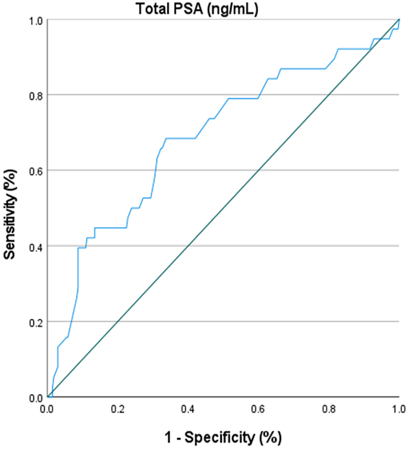
Figure 1. The receiver operating characteristic (ROC) curve illustrates the diagnostic accuracy of total prostate-specific antigen (PSA) in predicting lymph node invasion (LNI) in patients with intermediate-risk prostate cancer (PCa).
Discussion
Our findings revealed significant associations between LNI and several clinical factors in intermediate-risk PCa, including advanced clinical stage (T2c; OR=2.38), higher Gleason score (4+3; OR=2.64), increased core involvement (40-50%; OR=4.08; >50%; OR=8.16), and elevated total PSA levels (OR=1.33). Importantly, only PSA remained an independent predictor of LNI in multiple logistic regression analysis (OR=1.32 per ng/mL increase). ROC curve analysis confirmed the moderate diagnostic accuracy of total PSA for predicting LNI, demonstrating convergent findings with multiple previous investigations. 19 - 21 These results strongly aligned with established literature, where PSA has consistently emerged as the most robust independent predictor of lymph node metastasis. Porcaro and colleagues demonstrated that PSA was the sole independent predictor of bilateral LNI in high-risk PCa patients (OR=1.058), 20 while in another study, they confirmed PSA as the only independent predictor of extensive LNI with an OR of 1.054. 19
The percentage of positive cores, which showed strong univariate associations in our study, has been extensively validated as a leading predictor of LNI across multiple contemporary studies. Winter and colleagues found that the percentage of positive cores was the most accurate predictor of LNI, with 77% accuracy, surpassing PSA (71.1%) and clinical staging. 22 Briganti and colleagues also demonstrated that the percentage of positive cores was the most accurate predictor of LNI. 23 Recent large-scale analyses confirmed that patients with >50% positive cores demonstrated significantly elevated LNI rates. 24 , 25 A previous study showed that 91.4% of lymph node-positive patients had ≥50% core involvement. 25 Our observed Gleason score associations (4+3 pattern showing OR=2.64) were consistent with established risk stratification data, where intermediate-risk ISUP grade 3 patients demonstrated LNI rates of 5.1%, compared to 3.1-3.7% for those with lower grades. 11 Contemporary nomograms, including the extensively validated Briganti models, consistently identified PSA, clinical stage, Gleason grade, and percentage of positive cores as the fundamental predictors of LNI, with external validation studies confirming predictive accuracies ranging from 71-87.6%. 23 , 26 , 27 The singular independence of PSA in multiple logistic regression analysis, despite robust univariate associations for other factors, reflected the complex interdependence of PCa risk parameters and underscored PSA’s fundamental role as the most reliable preoperative predictor of lymph node metastasis across diverse patient populations and institutional settings.
The findings of the present study also revealed that factors, such as PCa heredity, age, BMI, prostate volume, and core involvement below 40%, demonstrated no statistically significant associations with LNI in intermediate-risk PCa patients. This finding that Pca heredity lacked significant association with LNI in intermediate-risk patients aligned with earlier evidence indicating that a positive family history did not independently predict nodal metastases. In a case-control study, self-reported family history—categorized as none, moderate (one affected first- or second-degree relative), or high (≥2 relatives)—showed no significant impact on tumor stage or lymph node involvement after radical prostatectomy (OR for moderate family history=1.48). 28 Similarly, patient’s age was shown not to influence LNI when other prognostic factors were controlled: a paired analysis across age groups (<55, 56–65, >65 years) demonstrated no differences in LNI rates among age-matched patients with equivalent PSA levels, Gleason scores, and biopsy involvement. 29
Contrasting with studies that report BMI as an independent predictor of multiple nodal metastases, 10 we found no significant BMI–LNI relationship. Notably, Briganti and colleagues observed that the frequency of positive lymph nodes did not differ across normal weight, overweight, and obese categories after extended PLND. 30 Likewise, although a study by Hong and colleagues linked larger prostate volume to increased LNI risk in univariate analysis, this association disappeared after adjusting for PSA, clinical stage, age, race, and BMI. 31 Collectively, these convergent findings underscored that heredity, age, BMI, and prostate volume did not confer additional predictive value for nodal metastasis beyond established factors, such as PSA, Gleason grade, clinical stage, and percentage of positive cores, and should not alter nodal staging strategies or the decision to perform extended PLND in intermediate-risk PCa patients.
First, the retrospective cross-sectional design of the study inherently limited the ability to establish causal relationships and might be subject to selection bias, as only patients who underwent radical prostatectomy with adequate lymph node dissection were included. Second, the study was conducted at a single tertiary care center, which might limit the generalizability of the findings to broader or more diverse populations. Third, the reliance on existing medical records and hospital information systems might have introduced information bias due to incomplete or inconsistent data documentation. Finally, intermediate-risk PCa was a heterogeneous group, and the study’s stratification might not fully capture the variability in tumor biology and behavior, potentially influencing the identification of risk factors associated with LNI. Future prospective multicenter studies with larger sample sizes are warranted to validate and extend these findings.
Conclusion
The findings of this study demonstrated that, in the unadjusted model, advanced clinical stage (T2c), higher Gleason score (4+3), and greater percentages of core involvement were significantly associated with increased risk of LNI in intermediate-risk PCa patients, alongside elevated total PSA levels. However, after adjusting for potential confounders, total PSA remained the only significant independent predictor of LNI, with each incremental increase in PSA conferring a higher risk. Although clinical stage T2c, Gleason score 4+3, and core involvement exceeding 50% indicated elevated ORs suggestive of increased LNI risk, these were not independent risk factors. Furthermore, ROC curve analysis confirmed the moderate diagnostic accuracy of total PSA for predicting LNI, with identified cut points balancing sensitivity and specificity. Collectively, these results underscore the central role of total PSA as an independent biomarker for lymph node involvement in intermediate-risk PCa, while other clinical and pathological factors might contribute to risk. However, they require further validation in adjusted models.
Acknowledgment
The study was supported by Tehran University of Medical Sciences (grant: 90713)
Authors’ Contribution
SA.M: Study design, data extraction, analysis, drafting, and reviewing the manuscript; M.A: Study design, and reviewing the manuscript, E.A: Statistical analysis, data curation, interpretation of results, and manuscript editing; SH.I: Statistical analysis, data curation, interpretation of results, and manuscript editing; MH.M: Study design, data interpretation, and reviewing the manuscript; EH.A: Data gathering, drafting, and reviewing the manuscript; F.GH: Data collection, data analysis, drafting, and reviewing the manuscript; MR.N: Conceptualization, data interpretation and reviewing the manuscript; and; All authors have read and approved the final manuscript and agree to be accountable for all aspects of the work in ensuring that questions related to the accuracy or integrity of any part of the work are appropriately investigated and resolved.
Declaration of AI
During the preparation of this work, the authors utilized AI (Perplexity.ai and Grammarly.com) to refine grammar points and language style in writing. Subsequently, the authors thoroughly reviewed and edited the content as necessary, assuming full responsibility for the publication’s content.
Conflicts of Interest
None declared.
References
- Ghaderi R, Abdollahi Z, Madani MH, Doshantapeh AG, Moghimi B, Jarang M, et al. Association between serum vitamin D levels and prostate tumor: a systematic review and meta-analysis. J Renal Inj Prev. 2024; 13:e34296. DOI
- Nourmohammadi A, A SA, K RA, B SQ, A RJ, Roohinezhad R, et al. The association between systemic immune-inflammation index and risk of prostate carcinoma; a systematic review and meta-analysis. Immunopathol Persa. 2025; 11:e43810. DOI
- Xu H, Zhu Y, Dai B, Ye DW. National Comprehensive Cancer Network (NCCN) risk classification in predicting biochemical recurrence after radical prostatectomy: a retrospective cohort study in Chinese prostate cancer patients. Asian J Androl. 2018; 20:551-4. Publisher Full Text | DOI | PubMed [ PMC Free Article ]
- Seiden B, Weng S, Sun N, Gordon D, Harris WN, Barnett J, et al. NCCN Risk Reclassification in Black Men with Low and Intermediate Risk Prostate Cancer After Genomic Testing. Urology. 2022; 163:81-9. DOI | PubMed
- Nezolosky M, Nguyen PL, Yang DD. Which patients with localized prostate cancer account for the greatest proportion of prostate cancer deaths? J Clin Oncol. 2018; 36:130. DOI
- Diven MA, Tshering L, Ma X, Hu JC, Barbieri C, McClure T, et al. Trends in Active Surveillance for Men With Intermediate-Risk Prostate Cancer. JAMA Netw Open. 2024; 7:e2429760. Publisher Full Text | DOI | PubMed [ PMC Free Article ]
- Harryman WL, Hinton JP, Sainz R, Gard JMC, Ryniawec JM, Rogers GC, et al. Intermediate risk prostate tumors contain lethal subtypes. Front Urol. 2024; 4Publisher Full Text | DOI | PubMed [ PMC Free Article ]
- Zumsteg ZS, Spratt DE, Pei I, Zhang Z, Yamada Y, Kollmeier M, et al. A new risk classification system for therapeutic decision making with intermediate-risk prostate cancer patients undergoing dose-escalated external-beam radiation therapy. Eur Urol. 2013; 64:895-902. DOI | PubMed
- Perera M, Lebdai S, Tin AL, Sjoberg DD, Benfante N, Beech BB, et al. Oncologic outcomes of patients with lymph node invasion at prostatectomy and post-prostatectomy biochemical persistence. Urol Oncol. 2023; 41:105. Publisher Full Text | DOI | PubMed [ PMC Free Article ]
- Tafuri A, Rizzetto R, Amigoni N, Sebben M, Shakir A, Gozzo A, et al. Predictors of Lymph Node Invasion in Patients with Clinically Localized Prostate Cancer Who Undergo Radical Prostatectomy and Extended Pelvic Lymph Node Dissection: The Role of Obesity. Urol Int. 2021; 105:362-9. DOI | PubMed
- Wenzel M, Preisser F, Hoeh B, Welte MN, Humke C, Wittler C, et al. Influence of Biopsy Gleason Score on the Risk of Lymph Node Invasion in Patients With Intermediate-Risk Prostate Cancer Undergoing Radical Prostatectomy. Front Surg. 2021; 8:759070. Publisher Full Text | DOI | PubMed [ PMC Free Article ]
- Maccio L, Barresi V, Domati F, Martorana E, Cesinaro AM, Migaldi M, et al. Clinical significance of pelvic lymph node status in prostate cancer: review of 1690 cases. Intern Emerg Med. 2016; 11:399-404. DOI | PubMed
- Hori S, Nakai Y, Tachibana A, Omori C, Nishimura N, Inoue K, et al. Clinical significance of limited and extended pelvic lymph node dissection during robot-assisted radical prostatectomy for patients with localized prostate cancer: A retrospective, propensity score matching analysis. Int J Urol. 2023; 30:168-75. DOI | PubMed
- Porcaro AB, de Luyk N, Corsi P, Sebben M, Tafuri A, Mattevi D, et al. Clinical Factors Predicting and Stratifying the Risk of Lymph Node Invasion in Localized Prostate Cancer. Urol Int. 2017; 99:207-14. DOI | PubMed
- Xu N, Ke ZB, Chen YH, Wu YP, Chen SH, Wei Y, et al. Risk Factors for Pathologically Confirmed Lymph Nodes Metastasis in Patients With Clinical T2N0M0 Stage Prostate Cancer. Front Oncol. 2020; 10:1547. Publisher Full Text | DOI | PubMed [ PMC Free Article ]
- Chen J, Wang Z, Zhao J, Zhu S, Sun G, Liu J, et al. Pelvic lymph node dissection and its extent on survival benefit in prostate cancer patients with a risk of lymph node invasion >5%: a propensity score matching analysis from SEER database. Sci Rep. 2019; 9:17985. Publisher Full Text | DOI | PubMed [ PMC Free Article ]
- Pollard ME, Hobbs AR, Kwon YS, Katsigeorgis M, Lavery HJ, Levinson A, et al. Heterogeneity of Outcomes in D’Amico Intermediate-Risk Prostate Cancer Patients after Radical Prostatectomy: Influence of Primary and Secondary Gleason Score. Oncol Res Treat. 2017; 40:508-14. DOI | PubMed
- Behmueller M, Tselis N, Zamboglou N, Zoga E, Baltas D, Rodel C, et al. High-Dose-Rate Brachytherapy as Monotherapy for Low- and Intermediate-Risk Prostate Cancer. Oncological Outcomes After a Median 15-Year Follow-Up. Front Oncol. 2021; 11:770959. Publisher Full Text | DOI | PubMed [ PMC Free Article ]
- Porcaro AB, Corsi P, Inverardi D, Sebben M, Tafuri A, Processali T, et al. Prostate-specific antigen associates with extensive lymph node invasion in high-risk prostate cancer. Tumori. 2018; 104:307-11. DOI | PubMed
- Porcaro AB, De Luyk N, Corsi P, Sebben M, Tafuri A, Processali T, et al. Clinical Factors Predicting Bilateral Lymph Node Invasion in High-Risk Prostate Cancer. Urol Int. 2017; 99:392-9. DOI | PubMed
- Wang X, Zhang X, Li H, Zhang M, Liu Y, Li X. Application of machine learning algorithm in prediction of lymph node metastasis in patients with intermediate and high-risk prostate cancer. J Cancer Res Clin Oncol. 2023; 149:8759-68. Publisher Full Text | DOI | PubMed [ PMC Free Article ]
- Winter A, Kneib T, Wasylow C, Reinhardt L, Henke RP, Engels S, et al. Updated Nomogram Incorporating Percentage of Positive Cores to Predict Probability of Lymph Node Invasion in Prostate Cancer Patients Undergoing Sentinel Lymph Node Dissection. J Cancer. 2017; 8:2692-8. Publisher Full Text | DOI | PubMed [ PMC Free Article ]
- Briganti A, Larcher A, Abdollah F, Capitanio U, Gallina A, Suardi N, et al. Updated nomogram predicting lymph node invasion in patients with prostate cancer undergoing extended pelvic lymph node dissection: the essential importance of percentage of positive cores. Eur Urol. 2012; 61:480-7. DOI | PubMed
- Shida Y, Hakariya T, Mitsunari K, Matsuo T, Ohba K, Miyata Y, et al. Preoperative Predictors of Lymph Node Invasion and Biochemical Recurrence in High-risk Prostate Cancer. Cancer Diagn Progn. 2022; 2:49-54. Publisher Full Text | DOI | PubMed [ PMC Free Article ]
- Heidenreich A, Pfister D, Thuer D, Brehmer B. Percentage of positive biopsies predicts lymph node involvement in men with low-risk prostate cancer undergoing radical prostatectomy and extended pelvic lymphadenectomy. BJU Int. 2011; 107:220-5. DOI | PubMed
- Gandaglia G, Martini A, Ploussard G, Fossati N, Stabile A, De Visschere P, et al. External Validation of the 2019 Briganti Nomogram for the Identification of Prostate Cancer Patients Who Should Be Considered for an Extended Pelvic Lymph Node Dissection. Eur Urol. 2020; 78:138-42. DOI | PubMed
- Briganti A, Capitanio U, Abdollah F, Gallina A, Suardi N, Bianchi M, et al. Assessing the risk of lymph node invasion in patients with intermediate risk prostate cancer treated with extended pelvic lymph node dissection. A novel prediction tool. Prostate 2012; 72:499-506. DOI | PubMed
- Spangler E, Zeigler-Johnson CM, Malkowicz SB, Wein AJ, Rebbeck TR. Association of prostate cancer family history with histopathological and clinical characteristics of prostate tumors. Int J Cancer. 2005; 113:471-4. DOI | PubMed
- da Cruz JAS, Passerotti CC, Dos Reis ST, Guariero MES, de Campos OD, Leite KRM, et al. Is Age an Independent Factor for Prostate Cancer? A Paired Analysis. Curr Urol. 2017; 9:183-7. Publisher Full Text | DOI | PubMed [ PMC Free Article ]
- Briganti A, Karakiewicz PI, Chun FK, Suardi N, Gallina A, Abdollah F, et al. Obesity does not increase the risk of lymph node metastases in patients with clinically localized prostate cancer undergoing radical prostatectomy and extended pelvic lymph node dissection. Int J Urol. 2009; 16:676-81. DOI | PubMed
- Hong SK, Poon BY, Sjoberg DD, Scardino PT, Eastham JA. Prostate size and adverse pathologic features in men undergoing radical prostatectomy. Urology. 2014; 84:153-7. Publisher Full Text | DOI | PubMed [ PMC Free Article ]

