
What’s Known
Pediatric Langerhans cell histiocytosis (LCH) commonly involves bone, especially the skull, and may present as lytic lesions with localized pain or swelling that can mimic traumatic or inflammatory conditions. Respiratory manifestations in LCH usually reflect pulmonary or airway involvement with objective abnormalities rather than isolated, reversible asthma-like symptoms.
What’s New
This case describes unifocal frontal bone Langerhans cell histiocytosis (LCH) presenting as persistent post-traumatic swelling, initially raising suspicion for a fracture or subgaleal hematoma, during the evaluation of an asthma exacerbation. The absence of clinical or chest radiographic pulmonary involvement supports interpreting the asthma exacerbation as a temporal overlap rather than a causal asthma–LCH relationship.
Introduction
Histiocytosis comprises a heterogeneous group of rare disorders characterized by clonal proliferation of myeloid-derived cells with phenotypic features of macrophages or dendritic cells. These pathological cells may infiltrate multiple organs, most commonly the skin, skeletal system, lungs, lymph nodes, and central nervous system. Clinical manifestations range from isolated, self-limited lesions to multisystem disease, with the prognosis largely determined by the extent and pattern of organ involvement.1, 2
Langerhans cell histiocytosis (LCH) has an estimated annual incidence of approximately 4.6 cases per million children younger than 15 years and shows a slight male predominance.3 Osseous involvement represents the most frequent manifestation in pediatric patients, with the skull being the most commonly affected site. Bone lesions typically demonstrate a lytic appearance on imaging and may be asymptomatic or associated with localized pain and soft tissue swelling, features that can complicate differentiation from traumatic or inflammatory conditions.4 The clinical course of LCH is highly variable and depends on the organs involved.
Although pulmonary involvement is a recognized feature of LCH, particularly in multisystem disease, respiratory manifestations usually reflect direct parenchymal or airway involvement. Asthma-like presentations have rarely been described in association with LCH, and reported cases generally involve persistent respiratory symptoms alongside objective pulmonary or airway abnormalities.
Published pediatric data suggest that respiratory symptoms are uncommon in children with pulmonary LCH and, when present, occur in patients with confirmed pulmonary disease. In a study reported by Wang and colleagues, only a small proportion of children with pulmonary LCH exhibited respiratory symptoms, despite objective pulmonary involvement characterized by findings such as cystic lung lesions and obstructive ventilatory abnormalities.5 In contrast, the present case involved transient respiratory symptoms attributed to an asthma exacerbation, which resolved following bronchodilator and corticosteroid therapy, with no subsequent persistent respiratory symptoms or clinical or chest radiographic evidence of pulmonary involvement. This contrast supports a cautious interpretation of the respiratory episode as a temporal clinical event rather than evidence of causality.
Within this context, we report a pediatric case in which a clinically diagnosed asthma exacerbation occurred during the diagnostic course of persistent post-traumatic frontal swelling, leading to the identification of unifocal osseous LCH in the absence of clinical or chest-radiographic pulmonary involvement, highlighting an unusual clinical presentation that may pose diagnostic challenges in routine practice.
Case Presentation
In 2022, a 4-year-old child from Guayaquil, Ecuador, was referred for evaluation of persistent frontal swelling following minor cranial trauma that had occurred approximately 1 month earlier. According to the mother, the child had struck his frontal region while playing with a yo-yo and subsequently developed painful frontal swelling initially presumed to be a post-traumatic hematoma (figure 1A). Initial home management with an oral nonsteroidal anti-inflammatory medication and topical treatment for bruising failed to produce clinical improvement; the lesion persisted without size reduction and remained painful. Approximately 1 week after the trauma, a conventional cranial computed tomography (CT) scan showed no acute intra-axial lesions, although it was initially reported as a depressed right frontal bone fracture with an associated subgaleal hematoma.
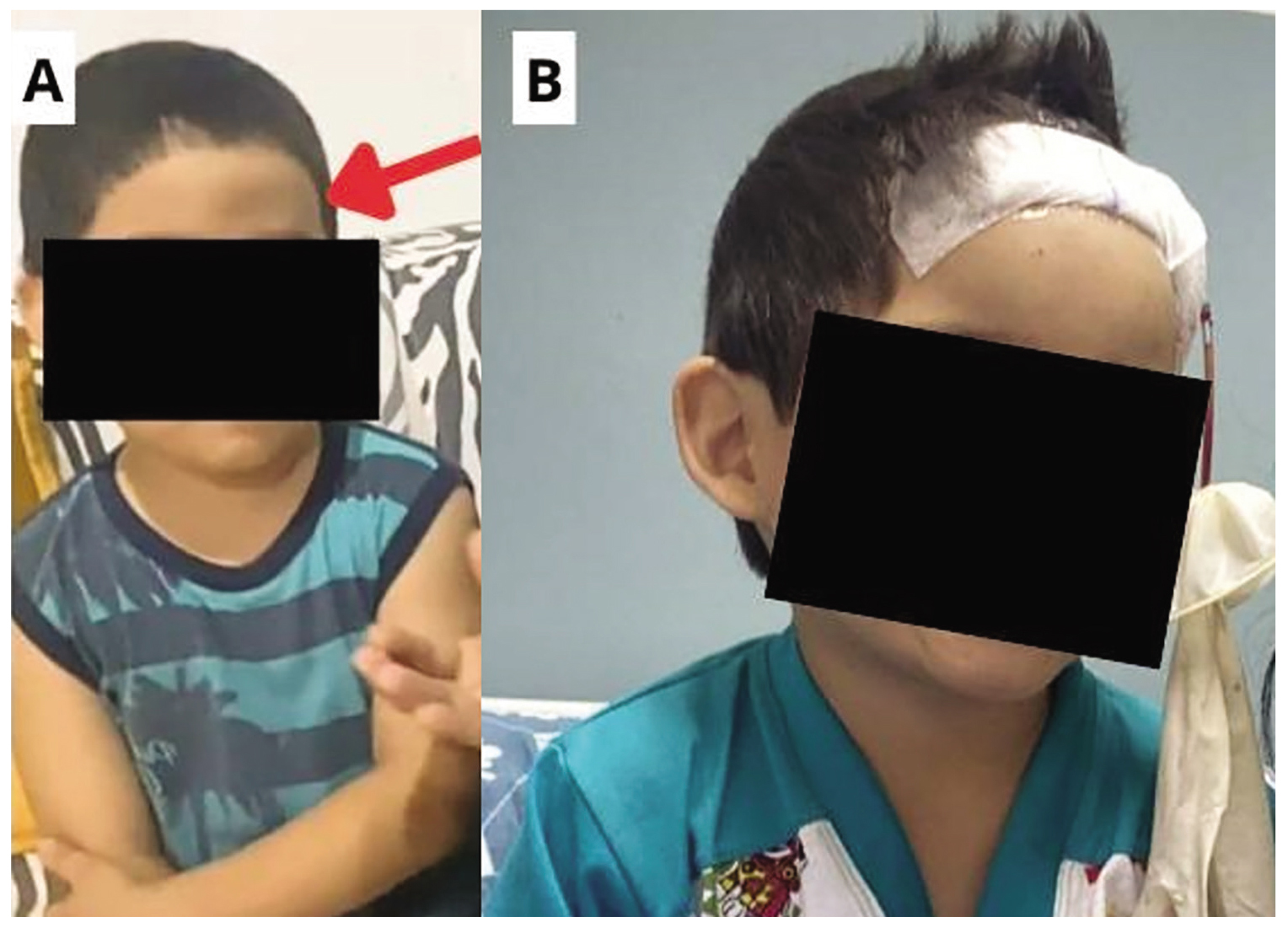
Figure 1.The clinical photographs show the frontal lesion before and after surgical management. A: It shows the preoperative right frontal swelling, which is indicated by a red arrow. B: It shows the postoperative appearance after surgical excision and curettage of the frontal skull lesion.
The patient had a history of recurrent respiratory illnesses during early childhood, including previous lower respiratory tract infections, with the last episode of pneumonia reported approximately 1 year before the current evaluation. Family history was notable for maternal asthma and atopic or allergic dermatitis in both parental lineages. Two weeks before the severe respiratory episode, the child had experienced a milder episode of wheezing and respiratory discomfort that resolved without hospitalization.
During the subsequent hospital evaluation, the child developed an acute, clinically diagnosed asthma exacerbation characterized by fever, respiratory distress, audible wheezing, intercostal and supraclavicular retractions, nasal flaring, and abnormal pulmonary auscultation. The episode was managed in the emergency department with repeated inhaled salbutamol every 20 min and systemic corticosteroid therapy, with clinical improvement. The patient was discharged with scheduled bronchodilator therapy for 1 week, with progressive spacing of doses.
At the cranial evaluation, physical examination revealed a firm, non-fluctuant, painful frontal mass. After treatment of the asthma exacerbation, no persistent respiratory symptoms, hypoxemia, or abnormal pulmonary findings were documented. Post-diagnostic upright posteroanterior and left lateral chest radiographs showed no findings suggestive of pulmonary involvement. Given the persistence of the lesion and its atypical evolution following minor trauma, further diagnostic evaluation was pursued. A skull X-ray demonstrated an area of osseous rarefaction with irregular margins in the right frontal bone (figure 2).
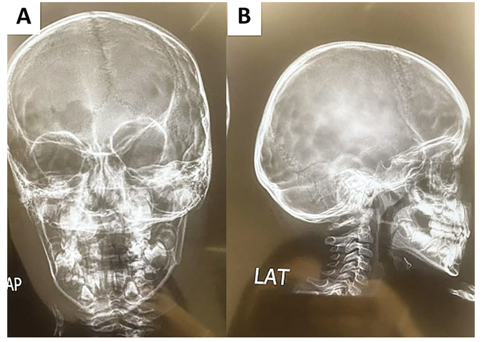
Figure 2.Skull radiography shows an area of bone rarefaction with irregular margins involving the right frontal bone. A: The image shows the anteroposterior view; B: It shows the lateral view.
Given that the respiratory episode was associated with fever and upper airway symptoms, a paranasal sinus CT scan was performed and showed paranasal sinus mucosal hypertrophy. Owing to the persistent painful frontal swelling and concern for an underlying cranial bone abnormality, a cranial CT scan with three-dimensional reconstruction was performed, which demonstrated a well-defined osteolytic lesion measuring approximately 4 cm in diameter in the frontal bone (figure 3).
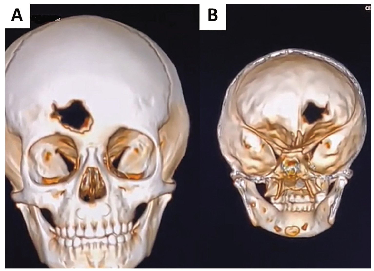
Figure 3.The three-dimensional reconstructed computed tomographic images demonstrate a well-defined osteolytic lesion measuring approximately 4 cm in diameter in the frontal bone. Panel A shows the external view, and Panel B shows the internal view.
Initial laboratory studies showed no evidence of systemic inflammation or hematologic abnormality, with a white blood cell count of 6.89×10⁹/L, hemoglobin of 11.4 g/dL, platelet count of 284,000/µL, and C-reactive protein of 0.02 mg/dL. Due to concern for an underlying pathological bone process, the patient was referred to a tertiary care center for specialized management. After multidisciplinary evaluation, the patient underwent surgical excision and curettage of the frontal skull lesion (figure 1B), and tissue samples were obtained for histopathological analysis.
Microscopic examination revealed features consistent with eosinophilic granuloma, including sheets of large polygonal cells with vesicular nuclei, prominent nuclear grooves, and pale eosinophilic cytoplasm, accompanied by scattered eosinophils and areas of fibrous tissue infiltration, confirming the diagnosis of LCH (figure 4). Based on the solitary frontal bone lesion and the absence of persistent respiratory symptoms, clinical pulmonary abnormalities, or chest radiographic findings suggestive of pulmonary involvement after treatment, the disease was classified clinically as single-system, unifocal osseous LCH involving the frontal bone, without clinical or chest radiographic evidence of pulmonary involvement.
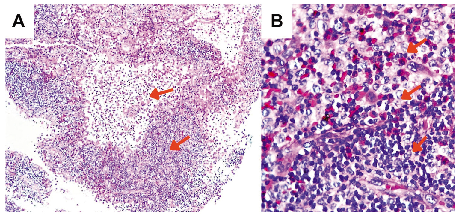
Figure 4.Histopathological examination of the excisional biopsy from the frontal osteolytic lesion shows findings consistent with Langerhans cell histiocytosis. Panel A shows loose to dense aggregates of polymorphous cells at low magnification (×100). Panel B shows large polygonal cells with vesicular nuclei, prominent nuclear grooves, and scattered eosinophils at high magnification(×400). Arrows indicate representative Langerhans-type cells with nuclear grooves and scattered eosinophils. Hematoxylin and eosin stain was used.
Following diagnosis, the patient was discharged and referred to pediatric oncology for outpatient chemotherapy. The medical record documented treatment with vinblastine 4.5 mg IV and prednisone 10 mg orally during the chemotherapy course. Commercial formulations were inferred from products available in Ecuador and were not specified in the medical record: vinblastine (Vinblastina Korea/Korea United Pharm Inc., South Korea) and prednisone (Meticorten/Schering-Plough, Ecuador listing). During treatment, routine laboratory monitoring demonstrated transient leukopenia, with a white blood cell (WBC) count of 2.8×10⁹/L and an absolute neutrophil count of 1,210/µL, requiring temporary suspension of chemotherapy. After hematologic recovery, chemotherapy was resumed, and follow-up laboratory studies showed recovery of hematologic parameters, including a WBC count of 5.52×10⁹/L, hemoglobin of 12.4 g/dL, platelet count of 391×109/L, and an absolute neutrophil count of 3,960/µL. The patient remained clinically stable and continued monthly outpatient follow-up in the pediatric oncology clinic for surveillance (table 1).
| Chronology | Variables | Clinical features and therapeutic approach |
|---|---|---|
| Day 0 | Minor frontal trauma | Frontal impact while playing with a yo-yo |
| Initial lesion | Painful frontal swelling initially presumed to be a post-traumatic hematoma. | |
| Days 1–14 | Initial home management | Oral nonsteroidal anti-inflammatory medication and topical treatment for bruising |
| Evolution | Persistent painful swelling without reduction in size | |
| Approximately days 7–14 after trauma | Conventional cranial CT | No acute intra-axial lesion; suspected depressed frontal fracture with subgaleal hematoma |
| Approximately 2 weeks before severe respiratory episode | Respiratory episode | Milder wheezing and respiratory discomfort, resolved without hospitalization. |
| Respiratory background | Previous history | Recurrent respiratory illnesses during early childhood, including previous lower respiratory tract infections |
| Previous pneumonia | Last pneumonia reported approximately 1 year before current evaluation. | |
| Family history | Maternal asthma and atopic/allergic dermatitis in both parental lineages | |
| Approximately 1 month after trauma/hospital evaluation | Severe respiratory episode | Fever, respiratory distress, audible wheezing, intercostal and supraclavicular retractions, nasal flaring, and abnormal pulmonary auscultation |
| Asthma management | Repeated inhaled salbutamol every 20 min and systemic corticosteroid therapy | |
| Outcome | Clinical improvement; discharged with scheduled bronchodilator therapy for one week with progressive spacing of doses | |
| Cranial evaluation | Physical examination | Firm, non-fluctuant, painful frontal mass |
| After asthma treatment | Pulmonary clinical status | No persistent respiratory symptoms, hypoxemia, or abnormal pulmonary findings |
| Post-diagnostic pulmonary evaluation | Chest radiographs | Upright posteroanterior and left lateral chest radiographs showed no findings suggestive of pulmonary involvement. |
| Chest CT | Not performed because there were no persistent respiratory symptoms, abnormal pulmonary examination, hypoxemia, or chest-radiographic abnormalities | |
| Cranial evaluation | Skull X-ray | Irregular bone rarefaction in the right frontal bone |
| Paranasal sinus CT | Paranasal sinus mucosal hypertrophy | |
| Cranial CT with 3D reconstruction | Well-defined osteolytic lesion measuring approximately 4 cm in the frontal bone | |
| Initial laboratory evaluation | White blood cell count | 6.890 cells/µL |
| Hemoglobin | 11.4 g/dL | |
| Platelets | 284.000 cells/µL | |
| C-reactive protein | 0.02 mg/dL | |
| Interpretation | No evidence of systemic inflammation or hematologic abnormality | |
| Hospital day 3 | Surgical intervention | Excision of frontal skull bone lesion and curettage of the frontal skull lesion |
| Biopsy sampling | Tissue sample sent for histopathological analysis. | |
| Histopathological evaluation | Histopathology | Large polygonal cells with irregular nuclei, nuclear grooves, eosinophilic cytoplasm, and scattered eosinophils, consistent with LCH/eosinophilic granuloma |
| Immunohistochemistry | CD1a (+), S-100 (+), Cyclin D1 (+), CD68 (+), CD21 (-) | |
| Systemic staging | Thyroid function tests | Within normal range |
| Abdominal ultrasound | Normal | |
| Bone marrow biopsy | Normocellular bone marrow without atypical cells; no bone marrow involvement identified | |
| Final clinical classification | Disease pattern | Single-system, unifocal osseous LCH involving the frontal bone, without clinical or chest-radiographic evidence of pulmonary involvement |
| Hospital discharge | Clinical status | Discharged and referred to pediatric oncology for outpatient chemotherapy |
| Week 1 of chemotherapy | Vinblastine | 4.5 mg IV |
| Prednisone | 10 mg orally | |
| Week 4 of chemotherapy | Vinblastine | 4.5 mg IV |
| Prednisone | 10 mg orally | |
| White blood cell count | 9.330 cells/µL | |
| Neutrophils | 78% | |
| Lymphocytes | 17% | |
| Absolute neutrophil count | 7.280 cells/µL | |
| Hemoglobin | 12.7 g/dL | |
| Platelets | 190.000 cells/µL | |
| C-reactive protein | 3.31 mg/dL | |
| Week 11 of chemotherapy | White blood cell count | 2.800 cells/µL |
| Absolute neutrophil count | 1.210 cells/µL | |
| Hemoglobin | 11.0 g/dL | |
| Hematocrit | 30% | |
| Platelets | 366.000 cells/µL | |
| Lactate dehydrogenase | 257 U/L | |
| Chemotherapy status | Temporarily suspended due to leukopenia. | |
| After hematologic recovery/final treatment week | Chemotherapy restart | Vinblastine 4.5 mg IV |
| White blood cell count | 5.520 cells/µL | |
| Absolute neutrophil count | 3.960 cells/µL | |
| Hemoglobin | 12.4 g/dL | |
| Platelets | 391.000 cells/µL | |
| Glucose | Within normal range | |
| Liver function tests | Within normal range | |
| Renal function | Within normal range | |
| Lactate dehydrogenase | 259 U/L | |
| Clinical outcome | Follow-up | Monthly outpatient pediatric oncology follow-up |
Written informed consent was obtained from the patient’s parents for the publication of this case report and the accompanying clinical images. Identifying information was removed to ensure patient anonymity.
Discussion
The present case described a 4-year-old child with LCH presenting as a solitary osteolytic lesion of the frontal bone, initially presumed to be a persistent post-traumatic hematoma. Imaging demonstrated an irregular lytic defect of the frontal bone, and histopathological examination confirmed eosinophilic granuloma consistent with osseous LCH. The diagnostic relevance of this case lies in the atypical persistence and progression of a seemingly benign cranial swelling. The asthma exacerbation that occurred during the diagnostic course added further clinical interest. However, its relationship with the subsequent diagnosis of LCH remained uncertain.
The post-traumatic presentation of the frontal swelling is also clinically relevant. Previous reports described skull eosinophilic granuloma/LCH becoming clinically apparent after minor head trauma.6 Although trauma should not be considered a proven cause of LCH, Kim and colleagues reported a rapidly growing skull eosinophilic granuloma after minor trauma, and Morimoto and colleagues described trauma-triggered bone lesions in a subset of pediatric patients with LCH.7 These observations supported the possibility that trauma might prompt medical attention, reveal a pre-existing lesion, or contribute to local inflammatory changes that make an underlying osteolytic lesion clinically evident, rather than directly causing LCH. Although the coexistence of minor trauma, a history of recurrent respiratory symptoms, an atopic background, and an asthma exacerbation is clinically notable, the available data did not support a unified triggering mechanism for LCH in this patient.
LCH is an uncommon disorder characterized by the clonal proliferation of dendritic cells, with osseous involvement representing the most frequent manifestation in pediatric patients. Although pulmonary disease is a recognized feature of LCH, respiratory manifestations usually reflect parenchymal or airway infiltration rather than isolated, reversible obstructive symptoms resembling asthma.
The temporal sequence observed in this patient is clinically noteworthy; nevertheless, it should be interpreted cautiously. Pediatric pulmonary involvement in LCH is uncommon and, when clinically apparent, is usually associated with persistent respiratory symptoms or objective pulmonary abnormalities. In a nationwide cohort of 1,482 children with LCH, lung involvement was identified in 111 patients (7.4%), whereas severe respiratory presentations requiring intensive care occurred in 17 patients (1.1%). These severe cases were associated with objective pulmonary abnormalities, including pneumothorax, extensive cystic lesions, or diffuse micronodular lung infiltration in the setting of multisystem disease.8
However, in the present case, the respiratory episode was clinically consistent with an asthma exacerbation in a preschool child with an atopic background and prior respiratory symptoms; it responded favorably to bronchodilators and was not followed by persistent symptoms or pulmonary involvement. Post-diagnostic two-view chest radiographs were unremarkable, with no findings suggestive of pulmonary involvement. Thus, the unusual feature was not merely that respiratory symptoms occurred before histopathological confirmation of LCH, but rather that a clinically diagnosed asthma exacerbation overlapped with the diagnostic course of solitary cranial bone LCH in the absence of either clinical or chest radiographic evidence of pulmonary involvement.
The significance of this temporal association remains uncertain. Given the patient’s respiratory history, family history of asthma and atopy, wheezing, increased work of breathing, and response to salbutamol and corticosteroids, the asthma exacerbation most likely represented an independent pediatric respiratory event. No causal or mechanistic relationship between the asthma exacerbation, the post-traumatic swelling, and the subsequent diagnosis of unifocal osseous LCH could be inferred from this single case.
To provide a more structured comparison with previously published cases, prior reports were interpreted according to three clinically relevant domains, including persistence of respiratory symptoms, presence of objective pulmonary or airway involvement, and the final pattern of LCH disease. Previously published reports further emphasized the distinction between this case and cases in which LCH mimicked asthma. In the pediatric case reported by Al-Trabolsi and colleagues, a 2-year-old child was initially treated for suspected asthma; however, the child subsequently developed acute respiratory failure, with radiographic findings consistent with pulmonary LCH. In that case, the respiratory compromise was attributable to direct pulmonary involvement rather than isolated airway hyperreactivity.9 In the adult case reported by Rawlins and colleagues, LCH presented as uncontrolled asthma. However, the clinical course was characterized by persistent respiratory symptoms that prompted further pulmonary evaluation and ultimately demonstrated objective pulmonary involvement.10 In contrast, our patient experienced a clinically diagnosed asthma exacerbation that resolved completely, with post-diagnostic chest radiographs showing no evidence of pulmonary involvement and the diagnosis of LCH confined to a solitary frontal bone lesion. This structured comparison supported the interpretation that the respiratory episode represented a temporal clinical overlap rather than evidence of a causal relationship between asthma and LCH.
This case underscored two clinically relevant points. First, persistent or progressively enlarging cranial swelling following minor trauma should not be assumed to represent a benign hematoma, particularly when it is associated with pain or demonstrates a progressive clinical course. Early imaging and histopathological confirmation are essential to exclude an underlying osteolytic condition such as LCH. Second, asthma exacerbations or asthma-like symptoms occurring during the diagnostic course of LCH should be interpreted cautiously when pulmonary involvement is present, as their coexistence might be incidental rather than disease-related. Recognizing this distinction is important to prevent both under-recognition of a rare systemic disorder and inappropriate attribution of common pediatric respiratory symptoms to LCH.
Conclusion
This case highlighted that persistent or painful cranial swelling after minor trauma should prompt early imaging and histopathological evaluation to exclude an underlying osteolytic disorder such as LCH. The clinically diagnosed asthma exacerbation occurred during the diagnostic course and represents a noteworthy temporal association with unifocal osseous LCH. However, no causal relationship between the respiratory episode and LCH could be inferred from this single case.
Authors’ Contribution
K.H. B-Z.: Conceptualization; data acquisition; data interpretation; manuscript drafting; critical revision; A.P.M: Data acquisition; clinical data review; manuscript revision; J.M.C: Clinical and imaging data acquisition; literature review; manuscript revision; A.V.C-C: Data organization; literature review; manuscript preparation; K.N.A.O: Clinical interpretation; pathology and imaging review; manuscript revision; A.D.B-Z: Literature review; data analysis support; manuscript editing; K.H.B-C: Supervision; critical revision for intellectual content; overall accountability. All authors have read and approved the final manuscript and agree to be accountable for all aspects of the work in ensuring that questions related to the accuracy or integrity of any part of the work are appropriately investigated and resolved.
Declaration of AI
The authors declare that no artificial intelligence tools, generative artificial intelligence systems, or AI-assisted technologies were used in the preparation, writing, editing, data analysis, interpretation, or revision of this manuscript.
Conflicts of Interest
None declared.
References
- Rocamora-Blanch G, Climent F, Solanich X. Histiocytosis. Med Clin (Barc). 2023; 161:166-75. DOI | PubMed
- Rodriguez-Galindo C, Allen CE. Langerhans cell histiocytosis. Blood. 2020; 135:1319-31. DOI | PubMed
- Allen CE, Merad M, McClain KL. Langerhans-Cell Histiocytosis. N Engl J Med. 2018; 379:856-68. Publisher Full Text | DOI | PubMed [ PMC Free Article ]
- Emile JF, Cohen-Aubart F, Collin M, Fraitag S, Idbaih A, Abdel-Wahab O. Histiocytosis. Lancet. 2021; 398:157-70. Publisher Full Text | DOI | PubMed [ PMC Free Article ]
- Wang D, Cui L, Li ZG, Zhang L, Lian HY, Ma HH. Clinical Research of Pulmonary Langerhans Cell Histiocytosis in Children. Chin Med J (Engl). 2018; 131:1793-8. Publisher Full Text | DOI | PubMed [ PMC Free Article ]
- Kim YJ, Jo KW. Rapid Growing Eosinophilic Granuloma in Skull after Minor Trauma. Korean J Neurotrauma. 2015; 11:22-5. Publisher Full Text | DOI | PubMed [ PMC Free Article ]
- Morimoto A, Shioda Y, Sakamoto K, Imamura T, Imashuku S, Japan LCHSG. Bone lesions of Langerhans cell histiocytosis triggered by trauma in children. Pediatr Int. 2022; 64:e15199. DOI | PubMed
- Le Louet S, Barkaoui MA, Miron J, Galambrun C, Aladjidi N, Chastagner P. Childhood Langerhans cell histiocytosis with severe lung involvement: a nationwide cohort study. Orphanet J Rare Dis. 2020; 15:241. Publisher Full Text | DOI | PubMed [ PMC Free Article ]
- Al-Trabolsi HA, Alshehri M, Al-Shomrani A, Shabanah M, Al-Barki AA. “Primary” pulmonary Langerhans cell histiocytosis in a two-year-old child: case report and literature review. J Pediatr Hematol Oncol. 2006; 28:79-81. DOI | PubMed
- Rawlins FA, Hull JE, Morgan JA, Morris MJ. Langerhans cell histiocytosis presenting as uncontrolled asthma. Case Rep Med. 2013; 2013:637232. Publisher Full Text | DOI | PubMed [ PMC Free Article ]


