
What’s Known
Anthracycline chemotherapy, despite improving breast cancer survival, is associated with dose-dependent cardiotoxicity that may lead to heart failure. Beta-blockers, especially carvedilol, indicated cardioprotective effects. However, evidence regarding bisoprolol remains scarce, and most studies have relied solely on ejection fraction.
What’s New
This triple-blind randomized controlled trial demonstrated that low-dose bisoprolol preserved left and right ventricular function during anthracycline-based chemotherapy. Using advanced echocardiographic parameters, the study revealed early cardioprotective effects of bisoprolol beyond conventional ejection fraction, addressing a significant gap in cardio-oncology research.
Introduction
Breast cancer is the most frequently diagnosed cancer worldwide. According to GLOBOCAN 2020 data, it accounted for approximately 2.3 million new cases (11.7% of new cancer cases). Furthermore, an estimated 685,000 women died from breast cancer in 2020, representing 16% of all cancer-related deaths in women.1, 2 Recently, cancer mortality has decreased significantly due to earlier diagnosis and the development of novel therapeutic modalities.3 Although many new-generation regimens have been developed, anthracyclines remain central components of breast cancer chemotherapy, reducing breast cancer-related mortality by up to 38%.3
However, despite improved cancer-free survival, complications of these therapies, particularly due to cardiac dysfunction, pose notable clinical challenges regarding long-term morbidity and mortality.3, 4 Anthracycline-induced cardiotoxicity (AIC) is a chronic, dose-dependent adverse effect that reduces left ventricular ejection fraction (LVEF) and may lead to cardiomyopathy, and in severe cases, heart failure (HF).5, 6 This cardiotoxicity is of significant clinical concern, as cardiovascular disease has become the leading cause of non-cancer-related mortality among breast cancer survivors.7, 8 This reality poses a significant therapeutic dilemma for oncologists, who must balance the oncologic efficacy of anthracyclines against their potential to cause lasting cardiac injury. In some cases, cardiotoxicity necessitates modification or discontinuation of potentially life-saving chemotherapy regimens.9
To address this issue, cardioprotective interventions are gaining increasing attention. Conventional strategies include limiting anthracycline dose, identifying high-risk patients, and early monitoring of cardiac function.10 In addition, pharmacologic prophylaxis has been explored as a means of mitigating cardiotoxic risk.11 Among these agents, beta-blockers, which have long been used in HF management, have demonstrated efficacy in preventing ventricular remodeling, reducing oxidative stress, and attenuating sympathetic overactivity, thus potentially offering cardioprotection during chemotherapy.12, 13
A variety of beta-blockers, such as metoprolol, atenolol, carvedilol, bisoprolol, and nebivolol, have been studied in this context.12, 14 Among these, carvedilol has received substantial attention, partly due to its antioxidant and anti-remodeling properties.15 Nevertheless, bisoprolol, a highly beta-1 selective agent with favorable tolerability, has also emerged as a potential prophylactic option in cardio-oncology. In a randomized trial, prophylactic bisoprolol was associated with attenuation of anthracycline-related declines in LVEF and global longitudinal strain (GLS) compared with placebo.16, 17
Despite these promising observations, there is a paucity of high-quality randomized trials specifically evaluating the cardioprotective role of bisoprolol in patients with breast cancer undergoing anthracycline therapy, particularly those utilizing comprehensive echocardiographic assessments. Previous studies have often relied on LVEF as the primary outcome, which might overlook early subclinical changes.16, 17 Therefore, in this study, we aimed to assess the cardioprotective effect of bisoprolol using a broad array of echocardiographic parameters, including left ventricular end-diastolic dimension (LVEDD), left ventricular end-systolic dimension (LVESD), left ventricular end-diastolic volume (LVEDV), GLS, tricuspid annular systolic excursion (TAPSE), and LVEF, to provide a more detailed evaluation of myocardial structural and functional integrity. We hypothesized that bisoprolol, when administered prophylactically during chemotherapy, could mitigate anthracycline-induced cardiotoxicity and preserve both left and right ventricular function in patients with breast cancer.
Materials and Methods
Study Design and Setting
This was a triple blind, randomized controlled trial conducted in Isfahan, Iran, between January and December 2023. Patients were consecutively recruited from the Oncology and Hematology Outpatient Clinics at Shariati Hospital, Isfahan, Iran. All patients were provided with oral and written information and provided their written informed consent prior to enrollment. The study protocol was approved by the Ethics Committee of Islamic Azad University, Najafabad Branch (code: IR.IAU.NAJAFABAD.REC.1400.058) and registered at the Iranian Registry of Clinical Trials (IRCT ID: IRCT20130701013828N8; Trial ID: 57932).
Population Selection
Inclusion criteria were adult female patients aged 18 to 70 years with newly diagnosed, histopathologically confirmed locally advanced breast cancer (Stage II or III), scheduled for anthracycline-based chemotherapy, and with a baseline LVEF ≥55%. To minimize selection bias, consecutive sampling was used. Each day, the research team screened all newly presenting patients using clinic records and admission lists. A structured screening form was completed to document demographic data, tumor characteristics, and planned chemotherapy regimen. Subsequently, a multidisciplinary team of oncologists and cardiologists reviewed each candidate to confirm eligibility based on predefined criteria.
Exclusion criteria included a history of cardiovascular disease (ischemic heart disease, HF, or arrhythmias), diabetes mellitus, chronic hypertension, or chronic kidney disease; previous exposure to chest radiotherapy or any form of chemotherapy; ongoing or planned use of beta-blockers for other medical indications; and any contraindication to bisoprolol administration, including acute decompensated HF, symptomatic bradycardia, or severe asthma. After confirmation of eligibility, detailed study information was provided, written informed consent was obtained, and baseline assessments, including physical examination and echocardiography, were performed. Randomization was carried out by blinded operators. Participant flow throughout the study is presented in the CONSORT diagram (figure 1).
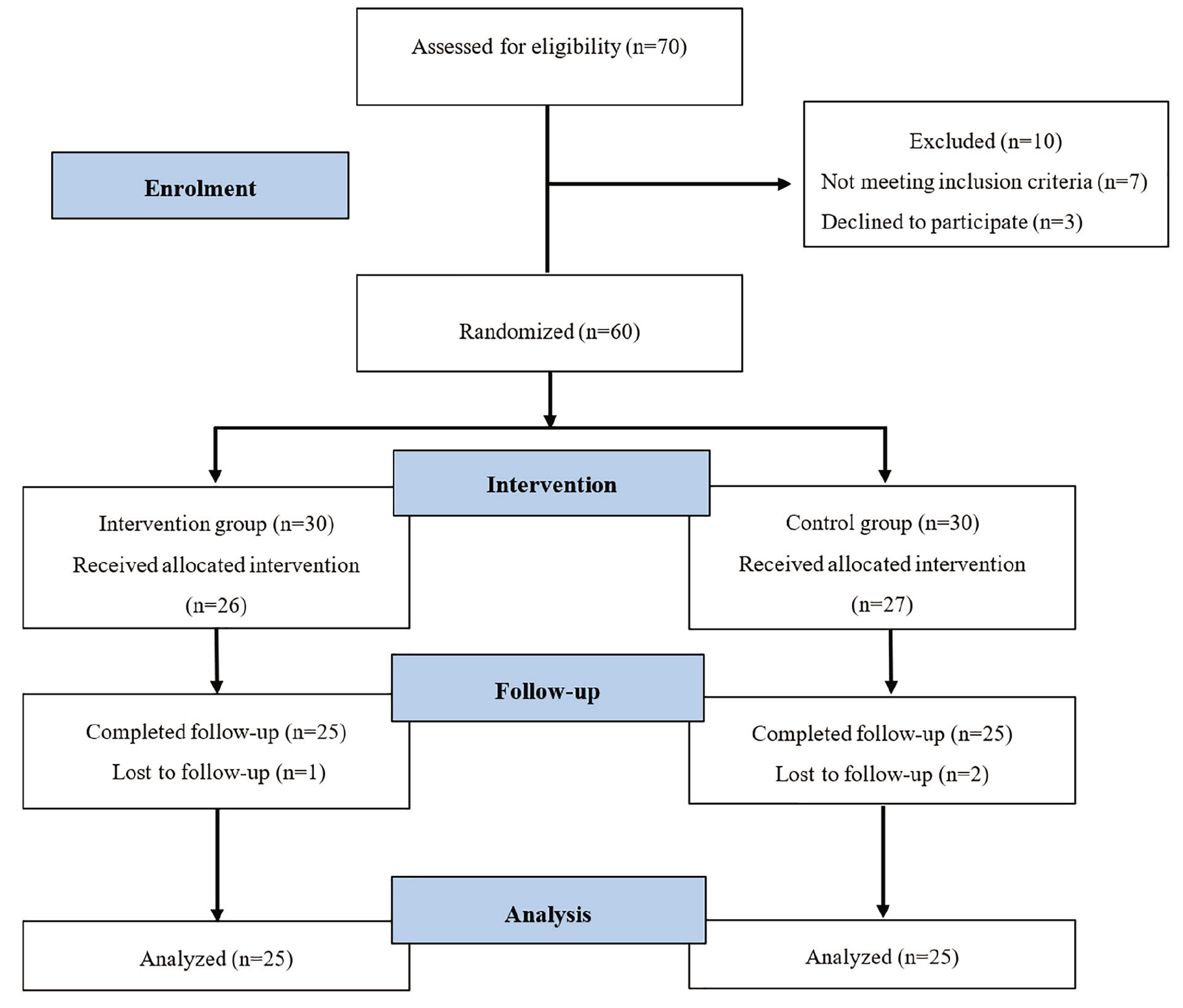
Figure 1.The CONSORT flow diagram shows the patient enrollment, randomization, intervention, follow-up, and analysis.
Randomization
Participants were randomized 1:1 to bisoprolol or placebo using a computer-generated block randomization sequence with a fixed block size (n=4) to maintain balanced group allocation. The randomization list was generated by an independent statistician using a random-number generator (SPSS software, version 25.0; IBM Corp., Armonk, NY, USA) and was not accessible to the investigators. The statistician encoded the allocation list and provided the treatment codes directly to the study pharmacist, who was responsible for preparing identical medication packages. Allocation concealment was ensured using sequentially numbered, opaque, sealed envelopes (SNOSE), which were opened only after eligibility confirmation.
Blinding Method
Patients were treated with medication that was prepared and packaged by an independent pharmacist, who was not involved in patient recruitment, treatment administration, or data analysis. Each package was identical in appearance, size, color, and labeling, regardless of whether it contained bisoprolol or placebo, to maintain effective blinding.
The study was conducted in a triple-blind manner: patients, clinical investigators responsible for patient management, and data analysts were all blinded to treatment allocation throughout the study period. Vital signs were monitored at each visit, and adverse events were managed per protocol without unblinding.
Study Treatment
All patients received in-hospital and outpatient chemotherapy according to our institution’s protocols. The regimens were as follows: cyclophosphamide (500-600 mg/m2)+epirubicin (75-CEF 100 mg/m2)+5-FU (500–600 mg/m2) (CEF regimen), or adriamycin (60 mg/m2)+cyclophosphamide (600 mg/m2) (AC regimen). All chemotherapy agents were administered as locally available commercial preparations and were identical between the two study groups. The intervention group received 2.5 mg of bisoprolol daily (manufactured by Abidi Pharmaceutical Co., Tehran, Iran, under the trade name of Betalco; n=25) and the control group received 2.5 mg of placebo daily (manufactured from pharmaceutical-grade microcrystalline cellulose provided by Abidi Pharmaceutical Co., Tehran, Iran; n=25). Placebo tablets were matched to the active drug for appearance, shape, weight, thickness, and color, and were packaged by an independent pharmacist to maintain blinding. The study drug was initiated orally at a dose of 2.5 mg for 7 consecutive days prior to chemotherapy. In the control group, placebo was also started 7 days before chemotherapy. Both bisoprolol and placebo were continued for 6 months.
Vital signs, including blood pressure and heart rate, were monitored before each chemotherapy cycle and monthly thereafter. If symptomatic bradycardia (heart rate<50 bpm) or hypotension (systolic BP<90 mmHg) occurred, the study drug was temporarily discontinued and permanently withdrawn upon recurrence.
Safety Monitoring
Adverse events were classified according to the Common Terminology Criteria for Adverse Events (CTCAE; version 5.0).18 In case of serious adverse events (Grade≥3), the safety committee was consulted, and emergency unblinding was permitted if necessary for patient safety.
Echocardiographic Assessments
Patients underwent transthoracic echocardiography at baseline, mid-treatment (at 3 months after chemotherapy initiation, approximately halfway through the anthracycline course), and end-of-treatment (at 6 months). If a reduction in LVEF ≥10% from baseline to a value <50% occurred, or if clinical signs of HF were observed, the study drug was stopped and guideline-directed medical therapy (GDMT), including initiation of angiotensin-converting enzyme inhibitors and beta-blockers (e.g., carvedilol), was started under cardio-oncology supervision. During follow-up, no participant met these criteria. Consequently, no patient required discontinuation of the study drug and initiation of GDMT.
Outcomes
The primary outcome of the study was the change in LVEF from baseline to following six cycles of chemotherapy. LVEF was measured using Simpson’s biplane method according to the guidelines of the American Society of Echocardiography. Secondary outcomes included the incidence of a ≥10% absolute reduction in LVEF to below 50%, the development of clinically overt HF classified by the New York Heart Association (NYHA) functional classification, changes in LVEDD and LVESD, changes in LVEDV, changes in myocardial deformation assessed by GLS, and changes in right ventricular systolic function evaluated by TAPSE. Echocardiographic studies were performed using the EPIC 7 ultrasound machine (Philips Medical Systems, Andover, MA, USA). All images were digitally stored and later analyzed offline directly on the echocardiography machine. All echocardiographic measurements were independently analyzed by an experienced echocardiography fellow.
Sample Size
The sample size was estimated to detect a clinically relevant 5-percentage-point difference in LVEF between the intervention and control groups. Based on previous cardio-oncology trials, a standard deviation (SD) of 5%, a two-sided α of 0.05, and 90% statistical power were assumed.16 Using the two-sample comparison of means formula, the minimum required sample size was calculated as 21 participants per group. Since patients receiving anthracycline chemotherapy commonly experience treatment discontinuation due to toxicity, clinical progression, withdrawal of consent, or loss to follow-up, an anticipated attrition rate of approximately 30% was incorporated. After adjustment for expected attrition, the target enrollment increased to 30 patients per group (total=60), ensuring that the final analyzable sample would meet the minimum powered requirement. To reach the target randomized sample of 60 patients, approximately 70 individuals were screened, accounting for the typical 10-20% exclusion rate observed during eligibility assessment in cardio-oncology trials.19 Reasons for pre-randomization exclusion include baseline cardiac abnormalities, previous chemotherapy exposure, or declining participation. Due to feasibility constraints and recruitment limitations during the study period, 25 participants per group (total=50) were ultimately enrolled and completed post-chemotherapy evaluation. This final sample size remained sufficient to preserve the statistical power requirement after accounting for observed attrition.
Statistical Analysis
All statistical analyses were performed using the SPSS software (version 25.0; IBM Corp., Armonk, NY, USA). Descriptive statistics were presented as numbers and percentages for categorical data, and mean±SD and range for numerical data. Within-group analyses were assessed using paired t tests. Normality assumptions for paired differences were evaluated using the Shapiro–Wilk test. When data met normality criteria, mean differences and 95% confidence intervals were reported. Multiple imputation was prespecified in the statistical analysis plan to address potential missing data. However, in the present study, no missing data were observed in either primary or secondary outcome variables. Therefore, multiple imputation was not required, and all analyses were performed using complete cases only. No adjustments for multiple comparisons were applied, given the exploratory nature of secondary outcomes. Analyses were primarily conducted based on the intention-to-treat principle, including all randomized patients according to their assigned groups. P<0.05 was considered statistically significant.
Results
Baseline Characteristics
A total of 50 female patients were enrolled and randomized equally to two groups (n=25 per group). The overall mean age was 47.7±9.7 years, ranging from 31 to 69 years. The mean age in the bisoprolol group was 48.2±9.4 years, while in the control group it was 47.3±10.1 years, with no statistically significant difference between the groups.
Comparison of Left Ventricular End-Diastolic Dimensions Between and Within Groups
At baseline and mid-treatment, LVEDD values were comparable between the groups, with no significant differences. However, at the final measurement (LVEDD3), the bisoprolol group exhibited a significantly lower mean value than the control group (P=0.049). Within-group comparisons demonstrated significant differences between LVEDD1 and LVEDD2 (P=0.028) and between LVEDD2 and LVEDD3 (P=0.001), whereas no significant difference was observed between LVEDD1 and LVEDD3. In the control group, all pairwise comparisons revealed statistically significant increases over time (table 1).
| Group | Comparison | LVEDD (mean±SD) | P value |
|---|---|---|---|
| Between-group | Baseline (LVEDD1) | Bisoprolol: 4.552±0.301 cm | 0.708 |
| Control: 4.524±0.216 cm | |||
| Mid-treatment (LVEDD2) | Bisoprolol: 4.700±0.285 cm | 0.162 | |
| Control: 4.600±0.206 cm | |||
| Final (LVEDD3) | Bisoprolol: 4.520±0.389 cm | 0.049 * | |
| Control: 4.692±0.157 cm | |||
| Bisoprolol | LVEDD1 vs. LVEDD2 | 4.552±0.301 vs. 4.700±0.285 | 0.028 * |
| LVEDD2 vs. LVEDD3 | 4.700±0.285 vs. 4.520±0.389 | 0.001 * | |
| LVEDD1 vs. LVEDD3 | 4.552±0.301 vs. 4.520±0.389 | 0.649 | |
| Control | LVEDD1 vs. LVEDD2 | 4.524±0.216 vs. 4.600±0.206 | 0.004 * |
| LVEDD2 vs. LVEDD3 | 4.600±0.206 vs. 4.692±0.157 | 0.005 * | |
| LVEDD1 vs. LVEDD3 | 4.524±0.216 vs. 4.692±0.157 | <0.001 * | |
| Between-group comparisons were analyzed using independent-sample t tests, and within-group comparisons were assessed using paired-sample t tests. Data are presented as mean±SD. Echocardiographic measures are presented as absolute values. *P<0.05 was considered statistically significant. LVEDD: Left ventricular end-diastolic diameter | |||
Comparison of Left Ventricular End-Systolic Dimensions Between and Within Groups
As shown in table 2, no significant differences were found between the groups at baseline left ventricular end-systolic dimensions (LVESD1) or mid-treatment (LVESD2). However, at the final evaluation (LVESD3), the bisoprolol group showed significantly lower values than the control group (P=0.029).
| Group | Comparison | LVESD (mean±SD) | P value |
|---|---|---|---|
| Between-group | Baseline (LVESD1) | Bisoprolol: 3.500±0.304 cm | 0.508 |
| Control: 3.448±0.243 cm | |||
| Mid-treatment (LVESD2) | Bisoprolol: 3.512±0.248 cm | 0.898 | |
| Control: 3.520±0.184 cm | |||
| Final (LVESD3) | Bisoprolol: 3.480±0.317 cm | 0.029* | |
| Control: 3.636±0.122 cm | |||
| Bisoprolol | LVESD1 vs. LVESD2 | 3.500±0.304 vs. 3.512±0.248 | 0.740 |
| LVESD2 vs. LVESD3 | 3.512±0.248 vs. 3.480±0.317 | 0.256 | |
| LVESD1 vs. LVESD3 | 3.500±0.304 vs. 3.480±0.317 | 0.619 | |
| Control | LVESD1 vs. LVESD2 | 3.448±0.243 vs. 3.520±0.184 | 0.039* |
| LVESD2 vs. LVESD3 | 3.520±0.184 vs. 3.636±0.122 | 0.002* | |
| LVESD1 vs. LVESD3 | 3.448±0.243 vs. 3.636±0.122 | 0.001* | |
| Between-group comparisons were analyzed using independent-sample t tests, and within-group comparisons were assessed using paired-sample t tests. Data are presented as mean±SD. Echocardiographic measures are presented as absolute values. *P<0.05 was considered statistically significant. LVESD: Left ventricular end-systolic diameter | |||
Within-group comparisons indicated no significant differences across time in the bisoprolol group. In contrast, the control group showed statistically significant progressive increases in LVESD values across all pairwise comparisons.
Comparison of Left Ventricular End-Diastolic Volume Between and Within Groups
As presented in table 3, there were no statistically significant differences between the two groups at LVEDV1 or LVEDV2. However, at the final measurement (LVEDV3), the bisoprolol group had significantly lower values than the control group (P=0.033). Within-group analysis showed that in the bisoprolol group, LVEDV decreased significantly from LVEDV2 to LVEDV3 (P=0.002), with no significant changes observed from LVEDV1 to LVEDV2 or from LVEDV1 to LVEDV3. In the control group, LVEDV increased significantly from LVEDV2 to LVEDV3 (P<0.001) and from LVEDV1 to LVEDV3 (P<0.001), with a non-significant trend from LVEDV1 to LVEDV2.
| Group | Comparison | LVEDV (mean±SD) | P value |
|---|---|---|---|
| Between-group | Baseline (LVEDV1) | Bisoprolol: 67.40±13.04 cc | 0.810 |
| Control: 68.20±10.19 cc | |||
| Mid-treatment (LVEDV2) | Bisoprolol: 68.20±14.20 cc | 0.619 | |
| Control: 69.92±9.60 cc | |||
| Final (LVEDV3) | Bisoprolol: 66.56±14.49 cc | 0.033* | |
| Control: 74.48±10.66 cc | |||
| Bisoprolol | LVEDV1 vs. LVEDV2 | 67.40±13.04 vs. 68.20±14.21 | 0.374 |
| LVEDV2 vs. LVEDV3 | 68.20±14.21 vs. 66.96±14.49 | 0.002* | |
| LVEDV1 vs. LVEDV3 | 67.40±13.04 vs. 66.96±14.49 | 0.459 | |
| Control | LVEDV1 vs. LVEDV2 | 68.20±10.19 vs. 69.92±9.60 | 0.060 |
| LVEDV2 vs. LVEDV3 | 69.92±9.60 vs. 74.48±10.66 | <0.001* | |
| LVEDV1 vs. LVEDV3 | 68.20±10.19 vs. 74.48±10.66 | <0.001* | |
| Between-group comparisons were analyzed using independent-samples t tests, and within-group comparisons were assessed using paired-samples t tests. Data are presented as mean±SD. Echocardiographic measures are presented as absolute values. *P<0.05 was considered statistically significant. LVEDV: Left ventricular end-diastolic volume | |||
Comparison of Left Ventricular Ejection Fraction Between and Within Groups
Table 4 demonstrates that no statistically significant differences were found in LVEF between the two groups at baseline (LVEF1), mid-treatment (LVEF2), or the final evaluation (LVEF3). Within-group comparisons indicated that LVEF values remained stable over time in both the bisoprolol and control groups, with no statistically significant differences.
| Group | Comparison | LVEF (mean±SD) | P value |
|---|---|---|---|
| Between-group | Baseline (LVEF1) | Bisoprolol: 59.20±1.87% | 0.720 |
| Control: 59.00±2.04% | |||
| Mid-treatment (LVEF2) | Bisoprolol: 59.00±2.50% | <0.999 | |
| Control: 59.00±2.04% | |||
| Final (LVEF3) | Bisoprolol: 59.20±2.36% | 0.731 | |
| Control: 59.40±1.65% | |||
| Bisoprolol | LVEF1 vs. LVEF2 | 59.20±1.87 vs. 59.00±2.50 | 0.574 |
| LVEF2 vs. LVEF3 | 59.00±2.50 vs. 59.20±2.36 | 0.327 | |
| LVEF1 vs. LVEF3 | 59.20±1.87 vs. 59.20±2.36 | <0.999 | |
| Control | LVEF1 vs. LVEF2 | 59.00±2.04 vs. 59.00±2.04 | <0.999 |
| LVEF2 vs. LVEF3 | 59.00±2.04 vs. 59.40±1.65 | 0.161 | |
| LVEF1 vs. LVEF3 | 59.00±2.04 vs. 59.40±1.65 | 0.161 | |
| Between-group comparisons were analyzed using independent-samples t tests, and within-group comparisons were assessed using paired-samples t tests. Data are presented as mean±SD. Echocardiographic measures are presented as absolute values. P<0.05 was considered statistically significant. LVEF: Left ventricular ejection fraction | |||
Comparison of Global Longitudinal Strain Between and Within Groups
As shown in table 5, no significant differences were observed in GLS1 or GLS2 between the groups. However, at the final assessment (GLS3), the bisoprolol group exhibited a significantly better strain value than the control group (P=0.033).
Within-group analysis revealed a significant improvement from GLS2 to GLS3 in the bisoprolol group (P=0.042). The control group showed significant changes from GLS1 to GLS2 (P=0.013) and from GLS1 to GLS3 (P<0.001).
| Group | Comparison | GLS (mean±SD) | P value |
|---|---|---|---|
| Between-group | Baseline (GLS1) | Bisoprolol: -18.24±2.18 | 0.424 |
| Control: -18.77±2.44 | |||
| Mid-treatment (GLS2) | Bisoprolol: -17.78±1.85 | 0.677 | |
| Control: -17.60±1.25 | |||
| Final (GLS3) | Bisoprolol: -18.12±1.69 | 0.033* | |
| Control: -17.15±1.42 | |||
| Bisoprolol | GLS1 vs. GLS2 | -18.24±2.18 vs. -17.78±1.85 | 0.111 |
| GLS2 vs. GLS3 | -17.78±1.85 vs. -18.12±1.69 | 0.042* | |
| GLS1 vs. GLS3 | -18.24±2.18 vs. -18.12±1.69 | 0.730 | |
| Control | GLS1 vs. GLS2 | -18.77±2.44 vs. -17.60±1.25 | 0.013* |
| GLS2 vs. GLS3 | -17.60±1.25 vs. -17.15±1.42 | 0.072 | |
| GLS1 vs. GLS3 | -18.77±2.44 vs. -17.15±1.42 | <0.001* | |
| Between-group comparisons were analyzed using independent-samples t tests, and within-group comparisons were assessed using paired-samples t tests. Data are presented as mean±SD. Echocardiographic measures are presented as absolute values. *P<0.05 was considered statistically significant. GLS: Global longitudinal strain | |||
Comparison of Tricuspid Annular Systolic Excursion Between and Within Groups
As detailed in table 6, TAPSE values at baseline (TAPSE1) and mid-treatment (TAPSE2) did not differ significantly between the two groups. However, at the final evaluation (TAPSE3), the bisoprolol group exhibited a significantly higher value than the control group (P<0.001).
| Group | Comparison | TAPSE (mean±SD) | P value |
|---|---|---|---|
| Between-group | Baseline (TAPSE1) | Bisoprolol: 2.10±0.16 cm | 0.598 |
| Control: 2.12±0.15 cm | |||
| Mid-treatment (TAPSE2) | Bisoprolol: 2.06±0.15 cm | 0.907 | |
| Control: 2.06±0.07 cm | |||
| Final (TAPSE3) | Bisoprolol: 2.11±0.11 cm | <0.001 * | |
| Control: 1.94±0.12 cm | |||
| Bisoprolol | TAPSE1 vs. TAPSE2 | 2.10±0.16 vs. 2.06±0.15 | 0.382 |
| TAPSE2 vs. TAPSE3 | 2.06±0.15 vs. 2.11±0.11 | 0.202 | |
| TAPSE1 vs. TAPSE3 | 2.10±0.16 vs. 2.11±0.11 | 0.664 | |
| Control | TAPSE1 vs. TAPSE2 | 2.12±0.15 vs. 2.06±0.07 | 0.040* |
| TAPSE2 vs. TAPSE3 | 2.06±0.07 vs. 1.94±0.12 | <0.001* | |
| TAPSE1 vs. TAPSE3 | 2.12±0.15 vs. 1.94±0.12 | <0.001* | |
| Between-group comparisons were analyzed using independent-sample t tests, and within-group comparisons were assessed using paired-sample t tests. Data are presented as mean±SD. Echocardiographic measures are presented as absolute values. *P<0.05 was considered statistically significant. TAPSE: Tricuspid annular plane systolic excursion | |||
Within-group comparisons showed no significant changes over time in the bisoprolol group, whereas the control group demonstrated significant declines from TAPSE1 to TAPSE2 (P=0.040), TAPSE2 to TAPSE3 (P<0.001), and TAPSE1 to TAPSE3 (P<0.001). A visual summary of the longitudinal comparison of echocardiographic parameters is presented in figure 2.
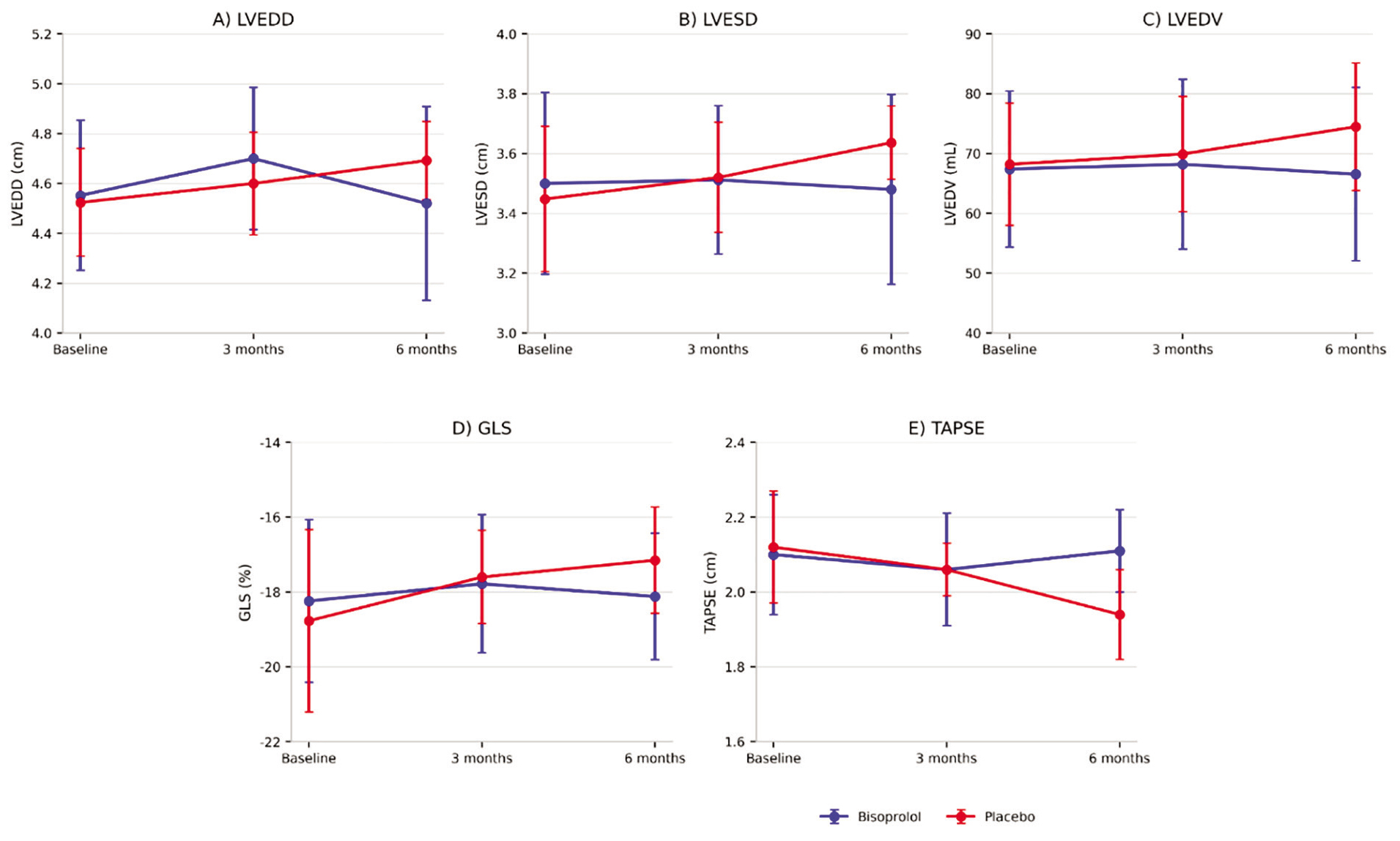
Figure 2.This figure shows longitudinal changes in echocardiographic parameters in the bisoprolol and placebo groups at baseline, 3 months, and 6 months: (A) LVEDD, (B) LVESD, (C) LVEDV, (D) GLS, and (E) TAPSE. Data are presented as mean±SD. Blue line indicates bisoprolol, and red line indicates placebo. LVEDD/LVESD/TAPSE are expressed in cm, LVEDV in mL, and GLS in %. LVEDD: Left ventricular end-diastolic diameter; LVESD: Left ventricular end-systolic diameter; LVEDV: Left ventricular end-diastolic volume; GLS: Global longitudinal strain; TAPSE: Tricuspid annular plane systolic excursion
Discussion
In this randomized controlled trial of female patients with breast cancer undergoing anthracycline-based chemotherapy, low-dose bisoprolol was associated with significant preservation of cardiac structure and function over time. While LVEF remained stable in both groups, the bisoprolol group showed significantly lower LVEDD, LVESD, and LVEDV at the final follow-up, improved GLS, and stable TAPSE than the declines in the control group. These findings supported the early initiation of bisoprolol for the prevention of anthracycline-induced cardiotoxicity and highlighted the need for preventive cardio-oncology strategies.
LVEDD is a sensitive echocardiographic index for detecting early geometric changes in the left ventricle, which often precede overt systolic dysfunction, especially in the context of anthracycline-induced cardiotoxicity.20 In this study, LVEDD was significantly lower in the bisoprolol group than the placebo group at 6 months. Nevertheless, the absolute between-group difference was small (≈2 mm), which may fall within routine echocardiographic measurement variability and may not represent a clinically meaningful change. Previous studies showed that anthracycline exposure was associated with progressive myocardial remodeling and dilation, reflected by increased LVEDD over time.21, 22 Our findings were broadly consistent with earlier reports, and a meta-analysis suggested beta-blockers, particularly carvedilol, might attenuate chemotherapy-related increases in LVEDD.14, 23 However, given the small magnitude, this finding should be interpreted cautiously and considered supportive rather than definitive.
LVESD is a key echocardiographic parameter that reflects systolic chamber size and indirectly indicates contractile function, with increases typically signaling progressive ventricular dysfunction.24 Previous studies emphasized the importance of LVESD as an early marker of subclinical systolic impairment during chemotherapy.21, 24 We observed statistically lower LVESD at final follow-up in the bisoprolol group than the placebo group. However, the absolute difference was small (2 mm), making its clinical impact uncertain in this short-term setting. Clinically, limiting LVESD progression is crucial given its association with later EF decline and HF.24 Accordingly, although the direction of effect is potentially favorable, larger trials with longer follow-up are required to determine whether such small early differences translate into meaningful clinical benefit.
LVEDV is a critical indicator of ventricular filling and compliance, and elevations in LVEDV often reflect early subclinical remodeling, particularly in patients exposed to anthracyclines.25 In this study, LVEDV remained essentially unchanged in the bisoprolol group, whereas patients in the placebo group experienced a progressive increase over time, resulting in significantly higher LVEDV at the final follow-up in those not receiving bisoprolol, which was consistent with the volumetric expansion associated with anthracycline-induced cardiotoxicity.25 These findings were in agreement with previous studies, such as meta-analyses by Shah and others, which demonstrated that beta-blockers attenuate left ventricular dilation and preserve cardiac geometry in patients undergoing chemotherapy.14 Stabilizing LVEDV might counter maladaptive volumetric expansion, reducing long-term HF risk.26-28
LVEF is a widely used measure of left ventricular systolic function in cardio-oncology,29 while in this study, LVEF remained stable across all time points in both the bisoprolol and control groups, with no statistically significant differences observed. Previous meta-analyses reported preserved LVEF in patients treated with beta-blockers despite anthracycline exposure, highlighting the limited sensitivity of LVEF in detecting early subclinical cardiotoxicity.14, 30, 31 While the preservation of LVEF might imply intact systolic function, it must be interpreted with caution since LVEF typically decreases only after considerable myocardial injury has occurred.32 Thus, favorable GLS and volume changes might better capture bisoprolol’s benefit even when LVEF appears stable.33
GLS has been identified as a sensitive echocardiographic marker for subclinical myocardial dysfunction at an early stage, particularly in patients undergoing anthracycline-based chemotherapy.34 In the present study, the divergence in GLS trajectories between the groups (improvement with bisoprolol versus worsening with placebo) represented one of the most clinically meaningful signals of early cardioprotection. These findings were consistent with previous research, which demonstrated the superiority of GLS over LVEF in early detection of cardiotoxicity and guiding intervention.35, 36 The U-shaped pattern observed in the bisoprolol group, with an initial non-significant decline followed by recovery, might reflect early myocardial adaptation mitigated by beta-blockade. However, this interpretation remains exploratory.37
TAPSE is an important echocardiographic index for assessing right ventricular (RV) systolic function, which can be impacted by anthracycline-induced cardiotoxicity, though it is often underexplored compared with LV function.38 However, similar to LVEDD and LVESD, the absolute difference was small (2 mm). Thus, the clinical relevance of this finding was uncertain over 6 months. These findings were in agreement with previous observations, which reported RV dysfunction following anthracycline therapy.39, 40 However, because TAPSE is load-dependent and reflects only a single component of RV mechanics, it should not be considered a comprehensive indicator of global RV performance.38 Thus, the between-group differences in TAPSE should be interpreted cautiously.
The strengths of this study were the triple-blind randomized design, standardized chemotherapy regimens, and the application of advanced echocardiographic parameters in addition to conventional indices for detection of early cardiac alterations. The three-point longitudinal evaluation enabled tracking of dynamic remodeling processes. However, limitations included a small sample size, a short follow-up period, restriction to female patients with breast cancer, the lack of comparison with other cardioprotective agents, a fixed low bisoprolol dose, potential interobserver variability in echocardiographic assessment, and the unavailability of body surface area-indexed parameters. These factors might affect generalizability and precision. Future work should confirm the cardioprotective effects of bisoprolol in large, multicenter, diverse cohorts with long-term follow-up, conduct head-to-head comparisons with other agents, and optimize dosing regimens.
Conclusion
This triple-blind trial suggested that low-dose bisoprolol during anthracycline chemotherapy in patients with breast cancer might mitigate early subclinical cardiotoxicity. LVEF remained stable in both groups. However, LVEDV was essentially preserved, and GLS evolved more favorably with bisoprolol, whereas LVEDV increased and GLS worsened in the placebo group. In contrast, the statistically significant changes in LVEDD, LVESD, and TAPSE were small and of uncertain clinical relevance. Larger, long-term studies are required to confirm its role in routine cardio-oncology care.
Acknowledgment
We thank the patients and clinical staff of the Oncology and Hematology Outpatient Clinics at Shariati Hospital (Isfahan, Iran) for their collaboration, and the Echocardiography Laboratory team for image acquisition and analysis. We would like to express our special gratitude to Dr. Mohammadreza Khosravi Farsani, Hematologist and Oncologist, for his valuable assistance in patient referral and support during the study. We are grateful to the independent statistician for generating the randomization sequence.
This article was derived from the MD thesis of Ms. Zahra Sadat Tabibian conducted at Islamic Azad University, Najafabad Branch, Iran. This research did not receive any specific grant from funding agencies in the public, commercial, or notforprofit sectors.
Authors’ Contribution
A.M: Coceptualization, study design, data interpretation, drafting and reviewing the manuscript; ZS.T: Dat gathering and drafting the manuscript, A.E: Data interpretation, drafting and reviewing the manuscript; HR.M: Data interpretation, drafting and reviewing the manuscript; All authors have read and approved the final manuscript and agree to be accountable for all aspects of the work in ensuring that questions related to the accuracy or integrity of any part of the work are appropriately investigated and resolved.
Declaration of AI
We acknowledge the use of ChatGPT (OpenAI) (version 5.4) for language polishing and paraphrasing during manuscript preparation; all content was reviewed, verified, and approved by the authors, who take full responsibility for the integrity and accuracy of the work.
Conflict of Interest
None declared.
References
- Ferlay J, Ervik M, Lam F, Colombet M, Mery L, Piñeros M. Global cancer observatory: cancer today. Lyon: International Agency for Research on Cancer; 2020.
- Cardinale D, Colombo A, Bacchiani G, Tedeschi I, Meroni CA, Veglia F. Early detection of anthracycline cardiotoxicity and improvement with heart failure therapy. Circulation. 2015; 131:1981-8. DOI | PubMed
- Jasra S, Anampa J. Anthracycline Use for Early Stage Breast Cancer in the Modern Era: a Review. Curr Treat Options Oncol. 2018; 19:30. DOI | PubMed
- Sobczuk P, Czerwinska M, Kleibert M, Cudnoch-Jedrzejewska A. Anthracycline-induced cardiotoxicity and renin-angiotensin-aldosterone system-from molecular mechanisms to therapeutic applications. Heart Fail Rev. 2022; 27:295-319. Publisher Full Text | DOI | PubMed [ PMC Free Article ]
- Gulati M, Mulvagh SL. The connection between the breast and heart in a woman: Breast cancer and cardiovascular disease. Clin Cardiol. 2018; 41:253-7. Publisher Full Text | DOI | PubMed [ PMC Free Article ]
- Dhir AA, Sawant SP. Cardiac morbidity & mortality in patients with breast cancer: A review. Indian J Med Res. 2021; 154:199-209. Publisher Full Text | DOI | PubMed [ PMC Free Article ]
- Li C, Dai H, Guo X, Zhou L, Jiang M. Comprehensive review of non-invasive-treatment-related cardiovascular toxicity in breast cancer. iScience. 2025; 28:111759. Publisher Full Text | DOI | PubMed [ PMC Free Article ]
- Cousin L, Roper N, Nolan TS. Cardio-Oncology Health Disparities: Social Determinants of Health and Care for Black Breast Cancer Survivors. Clin J Oncol Nurs. 2021; 25:36-41. Publisher Full Text | DOI | PubMed [ PMC Free Article ]
- Cho H, Lee S, Sim SH, Park IH, Lee KS, Kwak MH. Cumulative incidence of chemotherapy-induced cardiotoxicity during a 2-year follow-up period in breast cancer patients. Breast Cancer Res Treat. 2020; 182:333-43. DOI | PubMed
- Fabiani I, Chianca M, Cipolla CM, Cardinale DM. Anthracycline-induced cardiomyopathy: risk prediction, prevention and treatment. Nat Rev Cardiol. 2025; 22:551-63. DOI | PubMed
- Ponikowski P, Voors AA, Anker SD, Bueno H, Cleland JG, Coats AJ. [2016 ESC Guidelines for the diagnosis and treatment of acute and chronic heart failure]. Kardiol Pol. 2016; 74:1037-147. DOI | PubMed
- Curigliano G, Lenihan D, Fradley M, Ganatra S, Barac A, Blaes A. Management of cardiac disease in cancer patients throughout oncological treatment: ESMO consensus recommendations. Ann Oncol. 2020; 31:171-90. Publisher Full Text | DOI | PubMed [ PMC Free Article ]
- Blanter JB, Frishman WH. The Preventive Role of Angiotensin Converting Enzyme Inhibitors/Angiotensin-II Receptor Blockers and beta-Adrenergic Blockers in Anthracycline- and Trastuzumab-Induced Cardiotoxicity. Cardiol Rev. 2019; 27:256-9. DOI | PubMed
- Shah P, Garris R, Abboud R, Vasudev R, Patel H, Doshi R. Meta-Analysis Comparing Usefulness of Beta Blockers to Preserve Left Ventricular Function During Anthracycline Therapy. Am J Cardiol. 2019; 124:789-94. DOI | PubMed
- Avila MS, Ayub-Ferreira SM, de Barros Wanderley MR, das Dores Cruz F, Goncalves Brandao SM, Rigaud VOC. Carvedilol for Prevention of Chemotherapy-Related Cardiotoxicity: The CECCY Trial. J Am Coll Cardiol. 2018; 71:2281-90. DOI | PubMed
- Wihandono A, Azhar Y, Abdurahman M, Hidayat S. The Role of Lisinopril and Bisoprolol to Prevent Anthracycline Induced Cardiotoxicity in Locally Advanced Breast Cancer Patients. Asian Pac J Cancer Prev. 2021; 22:2847-53. Publisher Full Text | DOI | PubMed [ PMC Free Article ]
- Livi L, Barletta G, Martella F, Saieva C, Desideri I, Bacci C. Cardioprotective Strategy for Patients With Nonmetastatic Breast Cancer Who Are Receiving an Anthracycline-Based Chemotherapy: A Randomized Clinical Trial. JAMA Oncol. 2021; 7:1544-9. Publisher Full Text | DOI | PubMed [ PMC Free Article ]
- National Cancer Institute. Common terminology criteria for adverse events (CTCAE) version 5.0. Bethesda (MD): NCI; 2017.
- Unger JM, Vaidya R, Hershman DL, Minasian LM, Fleury ME. Systematic Review and Meta-Analysis of the Magnitude of Structural, Clinical, and Physician and Patient Barriers to Cancer Clinical Trial Participation. J Natl Cancer Inst. 2019; 111:245-55. Publisher Full Text | DOI | PubMed [ PMC Free Article ]
- Zhao R, Shu F, Zhang C, Song F, Xu Y, Guo Y. Early Detection and Prediction of Anthracycline-Induced Right Ventricular Cardiotoxicity by 3-Dimensional Echocardiography. JACC CardioOncol. 2020; 2:13-22. Publisher Full Text | DOI | PubMed [ PMC Free Article ]
- Moghadam AA, Fazaeli SH, Kamandi M. Left Ventricular Function and Echocardiographic Parameters in Patients Undergoing Chemotherapy With 5-Fluorouracil, Anthracycline, Taxotere, and Herceptin: Prospective Cohort Study. Health Sci Rep. 2025; 8:e71031. Publisher Full Text | DOI | PubMed [ PMC Free Article ]
- Nguyen KL, Hu P, Ennis DB, Shao J, Pham KA, Chen JJ. Cardiac MRI: a Translational Imaging Tool for Characterizing Anthracycline-Induced Myocardial Remodeling. Curr Oncol Rep. 2016; 18:48. Publisher Full Text | DOI | PubMed [ PMC Free Article ]
- Huang S, Zhao Q, Yang ZG, Diao KY, He Y, Shi K. Protective role of beta-blockers in chemotherapy-induced cardiotoxicity-a systematic review and meta-analysis of carvedilol. Heart Fail Rev. 2019; 24:325-33. Publisher Full Text | DOI | PubMed [ PMC Free Article ]
- Sandamali JAN, Hewawasam RP, Fernando M, Jayatilaka K, Madurawe RD, Sathananthan PP. Anthracycline-Induced Cardiotoxicity in Breast Cancer Patients from Southern Sri Lanka: An Echocardiographic Analysis. Biomed Res Int. 2020; 2020:1847159. Publisher Full Text | DOI | PubMed [ PMC Free Article ]
- Jordan JH, Sukpraphrute B, Melendez GC, Jolly MP, D’Agostino RB, Hundley WG. Early Myocardial Strain Changes During Potentially Cardiotoxic Chemotherapy May Occur as a Result of Reductions in Left Ventricular End-Diastolic Volume: The Need to Interpret Left Ventricular Strain With Volumes. Circulation. 2017; 135:2575-7. Publisher Full Text | DOI | PubMed [ PMC Free Article ]
- de Groote P, Delour P, Lamblin N, Dagorn J, Verkindere C, Tison E. Effects of bisoprolol in patients with stable congestive heart failure. Ann Cardiol Angeiol (Paris). 2004; 53:167-70. DOI | PubMed
- Ni Y, Wang T, Zhuo X, Song B, Zhang J, Wei F. Bisoprolol reversed small conductance calcium-activated potassium channel (SK) remodeling in a volume-overload rat model. Mol Cell Biochem. 2013; 384:95-103. DOI | PubMed
- Wang X, Zhao XZ, Wang XW, Cao LY, Lu B, Wang ZH. Effect of levosimendan on ventricular remodelling in patients with left ventricular systolic dysfunction: a meta-analysis. ESC Heart Fail. 2024; 11:1352-76. Publisher Full Text | DOI | PubMed [ PMC Free Article ]
- Stone JR, Kanneganti R, Abbasi M, Akhtari M. Monitoring for Chemotherapy-Related Cardiotoxicity in the Form of Left Ventricular Systolic Dysfunction: A Review of Current Recommendations. JCO Oncol Pract. 2021; 17:228-36. DOI | PubMed
- Attar A, Behnagh AK, Hosseini M, Amanollahi F, Shafiekhani P, Kabir A. Beta-Blockers for Primary Prevention of Anthracycline-Induced Cardiac Toxicity: An Updated Meta-Analysis of Randomized Clinical Trials. Cardiovasc Ther. 2022; 2022:8367444. Publisher Full Text | DOI | PubMed [ PMC Free Article ]
- Jeyaprakash P, Sangha S, Low G, Yu C, Pathan F, Negishi K. Prophylaxis to Prevent Cardiotoxicity in Patients Receiving Anthracycline for Breast Cancer: A Combined Bayesian and Frequentist Network Meta-Analysis of Randomised Controlled Trials. Heart Lung Circ. 2024; 33:710-20. DOI | PubMed
- Sutton NR, Li S, Thomas L, Wang TY, de Lemos JA, Enriquez JR. The association of left ventricular ejection fraction with clinical outcomes after myocardial infarction: Findings from the Acute Coronary Treatment and Intervention Outcomes Network (ACTION) Registry-Get With the Guidelines (GWTG) Medicare-linked database. Am Heart J. 2016; 178:65-73. DOI | PubMed
- Cocco LD, Chiaparini AF, Saffi MAL, Leiria TLL. Global Longitudinal Strain for the Early Detection of Chemotherapy-Induced Cardiotoxicity: A Systematic Review and Meta-analysis. Clin Oncol (R Coll Radiol). 2022; 34:514-25. DOI | PubMed
- Marwick TH. Global Longitudinal Strain Monitoring to Guide Cardioprotective Medications During Anthracycline Treatment. Curr Oncol Rep. 2022; 24:687-94. Publisher Full Text | DOI | PubMed [ PMC Free Article ]
- Stolojanu C, Steflea R, Micsescu-Olah AM, Alexandra I, Popoiu A, Doros G. Combined Utility of Speckle Tracking Echocardiography and Cardiac Biomarkers for Early Detection of Anthracycline-Induced Cardiotoxicity in Pediatric Oncology Patients. Biomedicines. 2024; 12Publisher Full Text | DOI | PubMed [ PMC Free Article ]
- Gonzalez-Manzanares R, Castillo JC, Molina JR, Ruiz-Ortiz M, Mesa D, Ojeda S. Automated Global Longitudinal Strain Assessment in Long-Term Survivors of Childhood Acute Lymphoblastic Leukemia. Cancers (Basel). 2022; 14Publisher Full Text | DOI | PubMed [ PMC Free Article ]
- Yurdam FS, Gurses E. Myocardial bridge and beta blockers: effect on left ventricular strain parameters. Acta Cardiol. 2023; 78:623-9. DOI | PubMed
- Aloia E, Cameli M, D’Ascenzi F, Sciaccaluga C, Mondillo S. TAPSE: An old but useful tool in different diseases. Int J Cardiol. 2016; 225:177-83. DOI | PubMed
- Abdar Esfahani M, Mokarian F, Karimipanah M. Alterations in the echocardiographic variables of the right ventricle in asymptomatic patients with breast cancer during anthracycline chemotherapy. Postgrad Med J. 2017; 93:271-4. DOI | PubMed
- Hu HM, Zhang XL, Zhang WL, Huang DS, Du ZD. Detection of Subclinical Anthracyclines’ Cardiotoxicity in Children with Solid Tumor. Chin Med J (Engl). 2018; 131:1450-6. Publisher Full Text | DOI | PubMed [ PMC Free Article ]


