
What’s Known
Monounsaturated fatty acids (MUFAs) are a key component of dietary fat, often considered beneficial, particularly in the context of the Mediterranean diet. Previous observational studies indicated conflicting results on the relationship between MUFA intake or serum levels and cardiovascular disease (CVD) risk. There is a lack of Mendelian randomization (MR) studies evaluating the association effect of serum MUFA levels (rather than dietary intake) on CVD outcomes.
What’s New
This Mendelian randomization (MR) study investigated the relationship between genetically predicted serum monounsaturated fatty acid (MUFA) levels and multiple cardiovascular disease (CVD) outcomes, such as angina, atherosclerosis, ischemic heart disease, myocardial infarction, and hypertension. The findings demonstrated that elevated serum MUFA levels were associated with an increased risk of these cardiovascular outcomes. The study suggested that endogenously elevated MUFA levels, rather than dietary intake alone, might play a role in CVD pathogenesis—providing new insights into metabolic contributions to cardiovascular risk.
Introduction
Fatty acids are a constituent of a group of carboxylic acids comprising saturated and unsaturated fatty acids. Saturated fatty acids lack double bonds, while monounsaturated fatty acids (MUFAs) and polyunsaturated fatty acids (PUFAs) have one double bond or more, respectively.1
The National Institutes of Health (NIH), the United States Department of Agriculture (USDA), the European Food and Safety Authority (EFSA), and the American Diabetes Association (ADA) have not provided appropriate guidelines regarding MUFA consumption. In contrast, the Academy of Nutrition and Dietetics recommends that 15%-20% of daily energy intake come from MUFAs,2 and the American Heart Association limits the consumption of MUFAs to 20% MUFA in their dietary guidelines.3 Additionally, dietitians and physicians highly recommend the Mediterranean diet for its health-promoting effects, which is rich in MUFAs.4
MUFAs may influence various markers associated with coronary heart disease (CHD), including postprandial vascular function, vascular function markers, and serum lipid and lipoprotein profiles.5 MUFA-enriched foods (e.g., plant-based oils) have also been recommended to reduce the risk of cardiovascular diseases (CVDs), body weight, and other inflammation-related diseases.6, 7 However, findings regarding the MUFA-CVD relationship are controversial, as some studies failed to find any significant association8-10 or reported a positive link.11
Previous studies examining the association between MUFA consumption and CVD risk have faced limitations and yielded inconclusive results.12-14 For instance, they mainly assessed dietary intake of MUFAs in relation to CVD events, whereas serum levels are a more precise indicator of MUFA consumption. The findings on the relationship between MUFA and CVD-related traits are predominantly based on observational studies, and the lack of clear insight is evident. Therefore, we aimed to explore whether high serum MUFA levels are an associated factor for CVD-related traits through a Mendelian randomization (MR) study.
Materials and Methods
This research report conformed to the strengthening the reporting of observational studies in epidemiology using Mendelian randomization (STROBE-MR) guidelines for observational studies using MR.15
Study Design
To estimate association inference, we selected serum levels of MUFAs as exposure and angina, atherosclerotic heart disease, ischemic heart disease (IHD), myocardial infarction (MI), and hypertension as outcomes from European ancestry (table 1, figure 1, and Supplementary figure S1).
| Name | GWAD ID | Sample size | Role |
|---|---|---|---|
| Angina | ukb-b-8468 | 461,880 (case=14,828, control=447,052) | Outcome |
| Atherosclerotic | ukb-b-1668 | 463,010 (case=12,171, control=450,839) | Outcome |
| Ischemic heart disease | ukb-d-I9_IHD | 361,194 (case=20,857, control=340,337) | Outcome |
| Myocardial infarction | ukb-b-15829 | 462,933 (case=10,616, control=452,317) | Outcome |
| High blood pressure | ukb-b-14177 | 461,880 (case=124,227, control=337,653) | Outcome |
| Monounsaturated fatty acids | met-d-MUFA | 114,999 | Exposure |
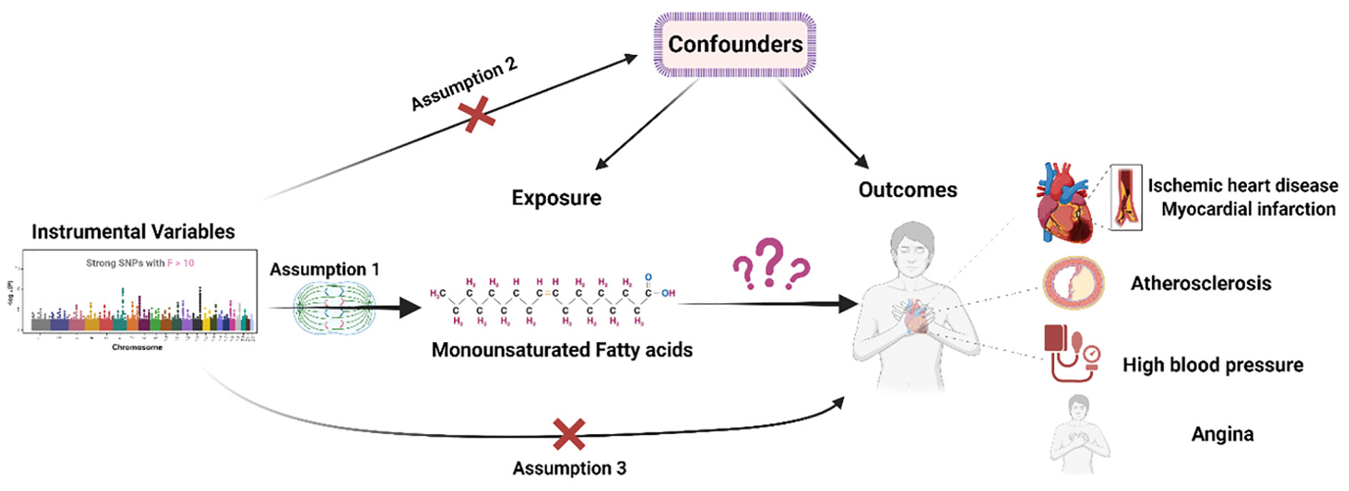
Figure 1.This figure illustrates the association between serum monounsaturated fatty acids and the risk of cardiovascular diseases, including angina, atherosclerotic heart disease, ischemic heart disease, myocardial infarction, and hypertension, based on Mendelian randomization analyses.
Generalized Summary Mendelian Randomization (GSMR)
GSMR is an extension of the summary-based Mendelian randomization (SMR) method. The SMR method requires individual-level data and employs a two-step least squares (2SLS) approach to estimate the effect of an exposure on an outcome. However, the statistical power of SMR can be restricted if the effect of the exposure on the outcome is small. GSMR, on the other hand, operates on summary-level data and can estimate the effect of an exposure on an outcome even if the effect is small.16
This method was developed according to previous approaches for modeling multiple correlated single-nucleotide polymorphisms (SNPs).17 Briefly, the process is extended to use all the top associated SNPs at a genome-wide significance level for the exposure as instrumental variables to test for causality.18 The statistical significance of the effect of the exposure on the outcome can be tested using a test statistic that follows a Chi squared distribution with 1 degree of freedom. This technique provides more power than the traditional MR approach because i) it accounts for the sampling variation in the estimated effects of the instruments on both the exposure and outcome, and ii) it accounts for linkage disequilibrium (LD) between the SNPs, whereas other methods assume independence among the instruments.19
We used the default settings in GSMR, including genome-wide significant SNPs with P<0.001 in common for the exposure, outcome, and the LD reference sample (1000 Genomes). Clump analysis was further performed with LD r 2 < 0.05 as instrumental variables in the GSMR analyses. Moreover, Heterogeneity in dependent instruments-outlier (HEIDI-outlier) detection analysis was implemented to test for horizontal pleiotropy (P<0.01) and remove SNPs with pleiotropic effects. An association relationship was declared significant at 0.05 divided by 5=0.001. For any associations, we also performed a TSMR.20
Two-Sample Mendelian Randomization (TSMR)
Genetic Variants of Exposure: We used summary GWAS information of the European ancestry on genetic variants associated with MUFAs and CVDs in figure 1. To specify the genetic instruments, we required a genome-wide threshold of P<0.001. Among all the identified variants, only SNPs were significantly associated with MUFAs at P<0.001. To minimize correlations between the SNPs, we conducted LD-clumping with a threshold of r 2=0.001, a clumping window of 10,000, and a European reference panel, in which SNPs with the lowest P value were retained.
For SNPs to be valid, they should be associated with the exposure, irrelevant to confounders of the exposure-outcome association, and should affect the outcome only through the exposure. The F-statistic was calculated to test the weak instrumental variable (IV) bias to avoid weak instrumental bias. If the F-statistic was greater than 10, the strength of the selected SNP was considered strong. We applied the PhenoScanner (http://www.phenoscanner.medschl.cam.ac.uk/) to assess whether the selected SNPs were associated with other traits at genome-wide significance levels. Then, after removing the identified pleiotropic SNPs, we repeated the analysis. The relevant information was extracted, including SNP, effect allele, non-effect allele, effect allele frequency, effect size, standard error, sample size, and P value.
Palindromic SNPs with intermediate allele frequencies (0.4-0.6) were disregarded from the above-selected SNPs. Additionally, SNPs with a minor allele frequency less than 0.01 were omitted to avoid potential statistical bias from the original GWAS, as they usually carry low confidence.
Statistical Analysis
The inverse variance-weighted (IVW) with a multiplicative random-effects model was performed to primarily analyze the associations between exposure to MUFAs and outcomes (CVDs). As this method might be affected by pleiotropy or invalid instrument bias, we assessed the validity and robustness of the results by executing several sensitivity analyses, including MR-Egger, weighted median, simple mode, and weighted mode methods. Moreover, we applied several other approaches, including the maximum likelihood estimation (MLE) method, robust adjusted profile score (RAPS), MR-Lasso, constrained maximum likelihood, mode-based, and debiased IVW methods to detect the robustness of our results. The MR Steiger test was performed to assess the direction of causation and whether the assumption that exposure causes the outcome was valid (P<0.05 was defined as statistically significant).
Cochran’s Q statistic and the I2 index for MR-IVW weighted analyses were used to detect heterogeneity and pleiotropy, and Rucker’s Q statistic was used for MR-Egger. We used the MR-Egger method (via intercept tests) to evaluate horizontal pleiotropy. Cook’s distance and Studentized residuals were applied to ascertain whether any individual SNPs were detected as outliers and influential points in driving the analysis results. Moreover, we used Mendelian randomization Pleiotropy Residual Sum and Outlier (MR-PRESSO) and RadialMR (using Cochran’s Q-statistic) to identify outliers with potential pleiotropy. To evaluate the validity and statistical power of the MR-Egger regression, we calculated the heterogeneity in instrument strength using the Q-statistic (weighted) and its derived I2-statistic (weighted). These metrics measure the variability in the associations between the genetic instruments and the exposure.
Leave-one-SNP-out analysis and its plot were conducted to assess the influence of potentially pleiotropic SNPs on the association estimates by systematically removing one SNP at a time. A funnel plot was depicted to detect directional pleiotropy, assessing whether association estimates from weaker variants tended to be skewed in one direction. We drew a forest plot of MR for the association between genetically predicted MUFAs and CVD. Additionally, we generated a scatter plot to visually inspect outliers, genetic association, and association estimates.
The principal statistical analyses were calculated with the help of R software (version 4.0.3; The R Foundation for Statistical Computing, Vienna, Austria) using the “TwoSampleMR” (MRC Integrative Epidemiology Unit [MRC IEU], University of Bristol, Bristol, UK) “MendelianRandomization” (MRC Biostatistics Unit, University of Cambridge, Cambridge, UK), “MR-PRESSO” (Icahn School of Medicine at Mount Sinai, New York, USA), “RadialMR” (MRC IEU, University of Bristol, Bristol, UK), and “mr. raps” (Harvard T.H. Chan School of Public Health, Boston, USA; developed by Qingyuan Zhao) packages. We also used the “mrrobust” package in Stata (MRC IEU, University of Bristol, Bristol, UK). Results were reported as odds ratios (ORs) with corresponding 95% confidence intervals (CIs). To account for multiple testing in MUFAs concerning the five outcomes, we used a Bonferroni-corrected threshold of P<0.01 (α=0.05/5 outcomes). Associations with P values between 0.01 and 0.05 were regarded as suggestive evidence of associations.
Ethics Approval and Consent to Participate
No additional ethics approval was required, as all data used in this study had been previously collected, analyzed, and published. The Isfahan University of Medical Sciences ethics committee approved this study (Research Approval Code: 340111; Research Ethical Code: IR.MUI.RESEARCH.REC.1401.063). Informed consent was obtained from all subjects in the original genome-wide association studies. The research was conducted in accordance with the Declaration of Helsinki.
Results
Generalized Summary Mendelian Randomization (GSMR)
After selecting SNPs in common among the exposure, outcome, the LD reference sample, and HEIDI-outlier analysis, results indicated that MUFAs had a positive association with angina, atherosclerotic heart disease, IHD, MI, and hypertension, as shown in figure 2.
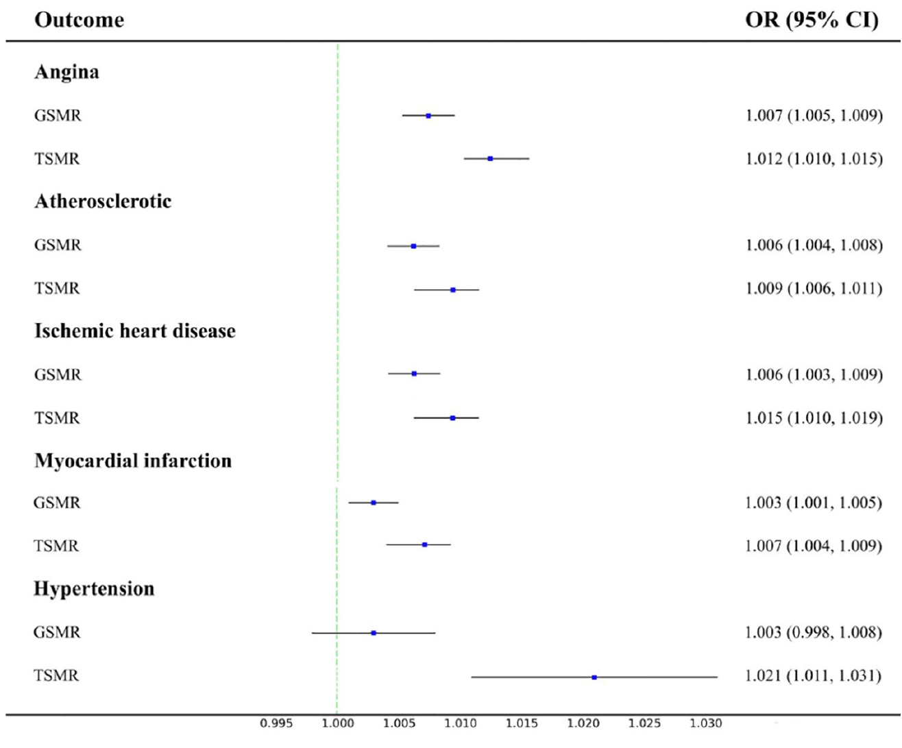
Figure 2.This figure presents the results of generalized summary Mendelian randomization and two-sample Mendelian randomization analyses, demonstrating a positive association between serum monounsaturated fatty acids and cardiovascular outcomes, including angina, atherosclerotic heart disease, ischemic heart disease, myocardial infarction, and hypertension. GSMR: Generalized summary Mendelian randomization; TSMR: Two-sample Mendelian randomization
Two-Sample Mendelian Randomization (TSMR)
SNP Selection: From 12,321,875 SNPs of MUFAs, we obtained 66 SNPs after P<5×10⁻⁸ and LD clumping (Supplementary data). All F-statistics of SNPs were greater than 10, which indicated no weak instrument bias (Supplementary data). In the harmonizing step (action=2), after removing SNPs due to palindromic structure, the PhenoScanner tool, and potential outliers and influential points, we retained 45, 43, 46, 41, and 46 SNPs for angina, atherosclerotic, IHD, MI, and hypertension, respectively (Supplementary data).
To display the relationship of the undefined causal variables to the CVD outcomes of the study, we used the beta of MUFA regressed on the undefined causal variable versus the CVD outcome. All coefficients indicated a positive association between MUFA and CVD (Supplementary figure S2).
Mendelian Randomization Analysis
The IVW method showed that a positive association could exist between MUFAs and angina, atherosclerotic heart disease, IHD, MI, and hypertension (figure 2). The average F-statistics for angina, atherosclerotic, IHD, MI, and hypertension (HTN) were 27.9, 31.8, 28.4, 30.1, and 28.6, respectively.
After assessing the heterogeneity, pleiotropy test, potential outliers, and influential SNPs according to Cook’s distance, the results for angina were: OR=1.012, 95% CI:1.010–1.015, P≤0.001; Cochran’s Q statistic=51.09, P=0.214; Rucker’s Q statistic=46.81, P=0.318; I2=8.2%, 95%CI:0.0%-36.2%; Q-statistic (weighted)=1579.38, I2-statistic (weighted)=97.21%; Egger intercept=0.0002, P=0.053.
For atherosclerotic heart disease, the results were: OR=1.009, 95% CI:1.006–1.011, P≤0.001; Cochran’s Q statistic=51.14, P=0.157; Rucker’s Q statistic=47.68, P=0.219; I2=14.0%, 95%CI: 0.0%-41.6%; Q-statistic (weighted)=1725.08, I2-statistic (weighted)=97.57%; Egger intercept= 0.0001, P=0.092.
For IHD, the results were: OR=1.015, 95% CI: 1.010-1.019, P ≤0.001; Cochran’s Q statistic=57.86, P=0.094; Rucker’s Q statistic=53.12, P=0.162; I2=17.2%, 95%CI: 0.0%-43.2%; Q-statistic (weighted)=978.06, I2-statistic (weighted)=95.40%; Egger intercept=0.0004, P=0.054.
For MI, the results were: OR=1.007, 95% CI:1.004–1.009, P≤0.001; Cochran’s Q statistic=39.12, P=0.510; Rucker’s Q statistic=38.10, P=0.511; I2=0.0%, 95%CI: 0.0%-36.2%; Q-statistic (weighted)=1080.45, I2-statistic (weighted)=96.30%; Egger intercept=0.0001, P=0.318.
For hypertension, the results were: OR=1.021, 95% CI:1.011–1.031, P≤0.001; Cochran’s Q statistic=86.02, P=2.101390e-05; Rucker’s Q statistic=87.78, P=1.964347e-05; I2=81.4%, 95%CI:75.8%, 85.7%; Q-statistic (weighted)=1455.39, I2-statistic (weighted)=96.91%; Egger intercept=0.0004, P=0.387.
Both MR analyses consistently demonstrated a positive association effect of higher serum MUFAs levels on the risk of several CVD outcomes. Genetically predicted MUFA levels were significantly associated with an increased risk of angina, atherosclerotic heart disease, IHD, MI, and hypertension. The findings were robust across different MR methods and sensitivity analyses.
Sensitivity Analyses
According to the results of Cochran’s Q test, statistically significant heterogeneity was observed only in hypertension. MR-Egger regression intercept analysis revealed no directional horizontal pleiotropy for any CVD (supplementary figure S3).
MR-PRESSO methods, Radial MR, and Cook’s distance were used to assess the presence of outlying SNPs and to reassess the effect estimates (supplementary figure S4). Densities of the IV estimates based on the mode method (supplementary figure S5), scatter plots (supplementary figure S6), and leave-one-out plots (supplementary figure S7) were visually examined to test the influence of outlying values.
Moreover, we depicted funnel and forest plots of the association between MUFAs and CVD diseases (supplementary figures S8 and S9). Weak instrumental variables were evaluated through the F-statistic (Supplementary Data).
We also checked whether SNPs within the HLA region existed in our MR analysis (Supplementary Data). Additionally, the MR Steiger test indicated that the correct association direction existed for CVDs (P<0.001). A summary of the sensitivity analysis can be found in figure 3 and supplementary figure S10.
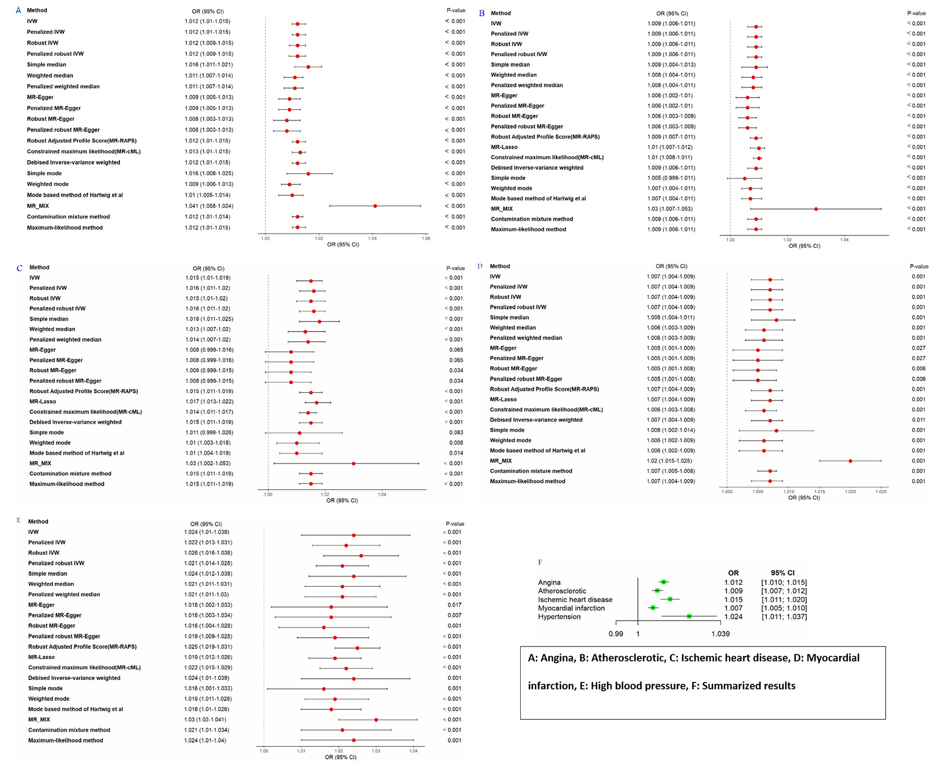
Figure 3.This figure displays the results of sensitivity analyses for the two-sample Mendelian randomization study, with panels A through E showing the effects of serum monounsaturated fatty acids on angina, atherosclerotic heart disease, ischemic heart disease, myocardial infarction, and hypertension, respectively, and panel F summarizing the overall effect of serum monounsaturated fatty acids on cardiovascular diseases. MR: Mendelian randomization; IVW: Inverse variance weighted; MR-MIX: Mendelian randomization analysis using mixture model; OR: Odds ratio; CI: Confidence interval
Discussion
The positive association between MUFAs and CVD events reported in this study was in line with the findings of several epidemiological studies.21-23 Consistent with the findings of the present study, a meta-analysis of five UK population-based cohorts and one matched case-control study suggested that high levels of circulating MUFAs, such as palmitoleic acid and oleic acid, were associated with a higher risk of coronary heart disease and stroke.21 Similarly, Akbaraly and others showed that higher levels of serum MUFAs were linked with low adherence to a healthy diet, reporting a positive association between elevated MUFA levels and increased CVD risk in the Whitehall II study population.22 Furthermore, an observational study conducted on CKD patients revealed that elevated serum MUFA levels were associated with an increased risk of CVDs.23 However, contrary to the results, a systematic review and meta-analysis reported a null association between dietary or circulating MUFAs and the risk of coronary diseases.24
Our findings were also comparable with results from prior research that explored the relationship between serum levels of individual MUFAs and CVD risk, which reported conflicting results. The prospective multi-ethnic study of atherosclerosis revealed that high circulating oleic acid levels might be a predictive risk factor for the incidence of CVD events and mortality.25 Likewise, high serum levels of palmitoleic acid were associated with an increased risk of ischemic stroke in a US cohort study,26 coronary heart disease,27 and coronary artery disease.28 In contrast, an inverse nonlinear association was found between linoleic acid and oleic acid, suggesting they might decrease risk of various CVD events, such as ischemic stroke,26 acute MI,29 and stroke incidence.30
In contrast with our findings, based on an MR analysis, Mazidi and others reported that genetically higher serum levels of some MUFAs were not associated with the risk of certain CVD events, such as coronary heart disease, MI, cardioembolic stroke, and ischemic stroke.10 One possible explanation for this controversy is that in the Mazidi and colleagues study, only some MUFAs were examined in relation to CVD risk, whereas the present study determined the association between total serum MUFAs and the risk of CVD events. It is noteworthy that a high total vascular concentration of these fatty acids can influence the connection between serum MUFA levels and CVD risk through their effects on vascular stiffness and susceptibility to CVD events. Identifying the relationship between serum MUFAs and these clinical abnormalities is more likely in our study, which focuses on total serum MUFA concentration. It is also important to note that the CVD outcomes examined in the present study differed from those assessed in the Mazidi and others study. The metabolic and biological mechanisms underlying CVDs might vary for different types of CVD events and, therefore, lead to different findings. Likewise, the direction and intensity of the effects of serum MUFAs on these various CVD events might differ.
Serum MUFAs are highly dynamic molecules that can influence a broad spectrum of cell signaling pathways related to lipid metabolism,21, 31 blood pressure regulation,32, 33 glucose homeostasis,34, 35 inflammation and oxidative stress,36 and endothelial function.37 MUFAs in the circulating fatty acid pool reflect the dynamic combination of several metabolic pathways, including adipose tissue lipolysis, re-esterification to triglycerides (TGs) within adipocytes, and peripheral utilization of fatty acids.21 Any unusual changes in these metabolic and biological pathways may have unfavorable metabolic and vascular consequences in the long term, particularly when the circulating levels of MUFAs are excessive. It is crucial to note that dietary intake of MUFAs does not have a significant role in determining serum MUFAs levels. Instead, endogenous synthesis is the primary factor in determining serum MUFA levels.21, 23 Therefore, these associations may indicate an underlying activation of hepatic de novo lipogenesis, which is linked to higher levels of blood TGs, abnormal fat deposition, and insulin resistance.38 Moreover, circulating fatty acid levels can affect several vital functions of the endothelium, such as regulating vascular tone, controlling blood flow and fluidity, modulating angiogenesis, and influencing inflammatory responses.37, 39, 40 Abnormal high levels of free fatty acids in the bloodstream can create an environment conducive to vascular dysfunction, encompassing a range of clinical disorders. Such vascular dysfunction can result in decreased nitric oxide production, increased cytokine production, impaired vasodilation, an overactive platelet response, escalated inflammation, and oxidative stress. The resulting clinical irregularities may ultimately lead to increased risk of CVD events.37
Although our findings indicated that high serum levels of MUFAs were causally linked to a greater risk of CVD outcomes, it is crucial to reiterate that these levels are primarily determined by endogenous metabolism rather than dietary MUFA intake.21, 23 Therefore, our results should not be interpreted as evidence for reducing the consumption of unsaturated fats. Instead, they highlighted the role of underlying metabolic processes, such as de novo lipogenesis, in influencing CVD risk. First, we did not determine the separate role of individual MUFAs in the blood in relation to cardiometabolic disorders. The biological role and metabolic pathways of each MUFA might differ, and this might influence serum levels of various types of MUFA subtypes and their impact on CVD outcomes. Moreover, different MUFAs may vary in their sensitivity and tendency to undergo metabolic processes, such as esterification, peroxidation, and calcification—factors that can affect their deposition and accumulation in the vessel wall. Therefore, recommending the consumption or restriction of one or more food groups that typically contain several types of unsaturated fatty acids might not be an effective intervention. Some MUFAs, such as palmitoleic acid and oleic acid, which are often present at high levels in the circulation and are frequently detected in biochemical measurements, do not necessarily reflect dietary levels of these fatty acids21, 23 as these fatty acids are also synthesized endogenously through de novo lipogenesis. Hence, limiting the consumption of food sources containing these fatty acids might not effectively reduce their serum concentrations.21, 23 These points might explain the inconsistency of our results with those of a previous meta-analysis, which reported that unsaturated fatty acids were not risk factors for CVD.24
The present study had important strengths. To our knowledge, this is the first study to use the TSMR method to assess the possible association between serum MUFAs and the risk of various CVD outcomes, providing a detailed and comprehensive view of the dose-response effect of serum MUFA levels on CVD risk. Another strength of the present study was the use of a large sample size from the UK Biobank database, which provided a rich source of genetic and phenotypic data. The assessment of cardiac function in clinical and research settings is multifaceted. While we utilized genetic proxies, other studies employed direct echocardiographic measures.41 However, some limitations should be acknowledged. The GWAS summary statistics for specific MUFAs—namely oleic acid and palmitoleic acid—were unavailable, preventing us from examining the association for each MUFA individually. Second, while the MR approach could suggest a causal relationship between serum MUFA levels and CVD risk, it did not elucidate the underlying mechanistic pathways. Experimental studies and clinical trials focusing on dietary MUFA intake are therefore required to clarify how dietary MUFAs influence serum MUFA levels and subsequently CVD risk. Finally, the genetic instruments and outcome data used in our analysis were derived exclusively from individuals of European ancestry. Although this approach reduced bias due to population stratification, it restricted the generalizability of our findings to other ethnic groups.
Conclusion
Evidence from our MR analysis supported a probable association between high serum levels of MUFAs and the risk of various CVD outcomes. However, these conclusions should be interpreted with caution due to the limitations of MR investigations and the complexity of the relationship between high serum MUFAs and CVD. Future studies involving populations with diverse characteristics, as well as the use of more precise analytical approaches and measurement tools, would be important to corroborate the evidence obtained from this study. Additionally, clinical studies are required to elucidate the underlying biological mechanisms that may explain the positive association observed between high serum MUFA levels and the risk of CVD events.
Acknowledgment
This article is derived from the doctoral dissertation of Danial Habibi.
Authors’ Contribution
DH: Data gathering, data analysis, interpretation, programming, and Software, writingoriginal draft; FT: Data gathering, writing original draft; FH: Data analysis, interpretation, verification, review and editing; STF: Data analysis, interpretation and reviewing the manuscript; SM: Data analysis, interpretation, invertigation, data curation and reviewing the manuscript; AST: Data analysis, interpretation, software and reviewing the manuscript; TR and SR: Data Curation, data analysis, interpretation and reviewing the manuscript; MR: Supervision, verification, review and editing; HF, HMM, HH, MH, MSD and AHS: Study concept, study design, data analysis, interpretation, and reviewing critically; FA: Study concept, review and editing; MM: Study concept, study design, supervision, review and editing. MA: Study concept, study design, data analysis, interpretation, data gathering, supervision, review, and editing. All authors have read and approved the final manuscript and agree to be accountable for all aspects of the work in ensuring that questions related to the accuracy or integrity of any part of the work are appropriately investigated and resolved.
Declaration of AI
The authors confirm that no AI-based tools were used in the conduct of this study or in the preparation of the manuscript.
Conflict of Interest
None declared.
References
- Albuquerque TG, Nunes MA, Bessada SMF, Costa HS, Oliveira MBPP. Chemical Analysis of Food. San Diego: Academic Press; 2020.
- Abdullah MM, Jew S, Jones PJ. Health benefits and evaluation of healthcare cost savings if oils rich in monounsaturated fatty acids were substituted for conventional dietary oils in the United States. Nutr Rev. 2017; 75:163-74. Publisher Full Text | DOI | PubMed [ PMC Free Article ]
- American Heart Association Nutrition C, Lichtenstein AH, Appel LJ, Brands M, Carnethon M, Daniels S. Diet and lifestyle recommendations revision 2006: a scientific statement from the American Heart Association Nutrition Committee. Circulation. 2006; 114:82-96. DOI | PubMed
- Mashek DG, Wu C. MUFAs. Adv Nutr. 2015; 6:276-7. Publisher Full Text | DOI | PubMed [ PMC Free Article ]
- Mika A, Sledzinski T. Alterations of specific lipid groups in serum of obese humans: a review. Obes Rev. 2017; 18:247-72. DOI | PubMed
- Hammad S, Pu S, Jones PJ. Current Evidence Supporting the Link Between Dietary Fatty Acids and Cardiovascular Disease. Lipids. 2016; 51:507-17. DOI | PubMed
- Rosqvist F, Niinisto S. Fats and oils - a scoping review for Nordic Nutrition Recommendations 2023. Food Nutr Res. 2024; 68Publisher Full Text | DOI | PubMed [ PMC Free Article ]
- Lotfi K, Salari-Moghaddam A, Yousefinia M, Larijani B, Esmaillzadeh A. Dietary intakes of monounsaturated fatty acids and risk of mortality from all causes, cardiovascular disease and cancer: A systematic review and dose-response meta-analysis of prospective cohort studies. Ageing Res Rev. 2021; 72:101467. DOI | PubMed
- Cheng P, Wang J, Shao W. Monounsaturated Fatty Acid Intake and Stroke Risk: A Meta-analysis of Prospective Cohort Studies. J Stroke Cerebrovasc Dis. 2016; 25:1326-34. DOI | PubMed
- Mazidi M, Katsiki N, Shekoohi N, Banach M. Monounsaturated Fatty Acid Levels May Not Affect Cardiovascular Events: Results From a Mendelian Randomization Analysis. Front Nutr. 2020; 7:123. Publisher Full Text | DOI | PubMed [ PMC Free Article ]
- Kim Y, Je Y, Giovannucci EL. Association between dietary fat intake and mortality from all-causes, cardiovascular disease, and cancer: A systematic review and meta-analysis of prospective cohort studies. Clin Nutr. 2021; 40:1060-70. DOI | PubMed
- Praagman J, Beulens JW, Alssema M, Zock PL, Wanders AJ, Sluijs I. The association between dietary saturated fatty acids and ischemic heart disease depends on the type and source of fatty acid in the European Prospective Investigation into Cancer and Nutrition-Netherlands cohort. Am J Clin Nutr. 2016; 103:356-65. DOI | PubMed
- Zong G, Li Y, Wanders AJ, Alssema M, Zock PL, Willett WC. Intake of individual saturated fatty acids and risk of coronary heart disease in US men and women: two prospective longitudinal cohort studies. BMJ. 2016; 355:i5796. Publisher Full Text | DOI | PubMed [ PMC Free Article ]
- Li Y, Hruby A, Bernstein AM, Ley SH, Wang DD, Chiuve SE. Saturated Fats Compared With Unsaturated Fats and Sources of Carbohydrates in Relation to Risk of Coronary Heart Disease: A Prospective Cohort Study. J Am Coll Cardiol. 2015; 66:1538-48. Publisher Full Text | DOI | PubMed [ PMC Free Article ]
- Skrivankova VW, Richmond RC, Woolf BAR, Yarmolinsky J, Davies NM, Swanson SA. Strengthening the Reporting of Observational Studies in Epidemiology Using Mendelian Randomization: The STROBE-MR Statement. JAMA. 2021; 326:1614-21. DOI | PubMed
- Zhu Z, Zheng Z, Zhang F, Wu Y, Trzaskowski M, Maier R. Causal associations between risk factors and common diseases inferred from GWAS summary data. Nat Commun. 2018; 9:224. Publisher Full Text | DOI | PubMed [ PMC Free Article ]
- Burgess S, Scott RA, Timpson NJ, Davey Smith G, Thompson SG, Consortium E-I. Using published data in Mendelian randomization: a blueprint for efficient identification of causal risk factors. Eur J Epidemiol. 2015; 30:543-52. Publisher Full Text | DOI | PubMed [ PMC Free Article ]
- Ong JS, MacGregor S. Implementing MR-PRESSO and GCTA-GSMR for pleiotropy assessment in Mendelian randomization studies from a practitioner’s perspective. Genet Epidemiol. 2019; 43:609-16. Publisher Full Text | DOI | PubMed [ PMC Free Article ]
- Jin C, Lee B, Shen L, Long Q, Alzheimer’s Disease Neuroimaging I, Alzheimer’s Disease Metabolomics C. Integrating multi-omics summary data using a Mendelian randomization framework. Brief Bioinform. 2022; 23Publisher Full Text | DOI | PubMed [ PMC Free Article ]
- Revez JA, Lin T, Qiao Z, Xue A, Holtz Y, Zhu Z. Genome-wide association study identifies 143 loci associated with 25 hydroxyvitamin D concentration. Nat Commun. 2020; 11:1647. Publisher Full Text | DOI | PubMed [ PMC Free Article ]
- Borges MC, Schmidt AF, Jefferis B, Wannamethee SG, Lawlor DA, Kivimaki M. Circulating Fatty Acids and Risk of Coronary Heart Disease and Stroke: Individual Participant Data Meta-Analysis in Up to 16 126 Participants. J Am Heart Assoc. 2020; 9:e013131. Publisher Full Text | DOI | PubMed [ PMC Free Article ]
- Akbaraly T, Wurtz P, Singh-Manoux A, Shipley MJ, Haapakoski R, Lehto M. Association of circulating metabolites with healthy diet and risk of cardiovascular disease: analysis of two cohort studies. Sci Rep. 2018; 8:8620. Publisher Full Text | DOI | PubMed [ PMC Free Article ]
- Mika A, Sikorska-Wisniewska M, Malgorzewicz S, Stepnowski P, Debska-Slizien A, Sledzinski T. Potential contribution of monounsaturated fatty acids to cardiovascular risk in chronic kidney disease. Pol Arch Intern Med. 2018; 128:755-63. DOI | PubMed
- Chowdhury R, Warnakula S, Kunutsor S, Crowe F, Ward HA, Johnson L. Association of dietary, circulating, and supplement fatty acids with coronary risk: a systematic review and meta-analysis. Ann Intern Med. 2014; 160:398-406. DOI | PubMed
- Steffen BT, Duprez D, Szklo M, Guan W, Tsai MY. Circulating oleic acid levels are related to greater risks of cardiovascular events and all-cause mortality: The Multi-Ethnic Study of Atherosclerosis. J Clin Lipidol. 2018; 12:1404-12. Publisher Full Text | DOI | PubMed [ PMC Free Article ]
- Yamagishi K, Folsom AR, Steffen LM, Investigators AS. Plasma fatty acid composition and incident ischemic stroke in middle-aged adults: the Atherosclerosis Risk in Communities (ARIC) Study. Cerebrovasc Dis. 2013; 36:38-46. Publisher Full Text | DOI | PubMed [ PMC Free Article ]
- Messner T, Sihm H, Vessby B. Fatty acid composition in serum among males 4-16 years after myocardial infarction. Int J Circumpolar Health. 1998; 57:22-31. PubMed
- Chei CL, Yamagishi K, Kitamura A, Kiyama M, Sankai T, Okada T. Serum Fatty Acid and Risk of Coronary Artery Disease - Circulatory Risk in Communities Study (CIRCS). Circ J. 2018; 82:3013-20. DOI | PubMed
- Lopes C, Aro A, Azevedo A, Ramos E, Barros H. Intake and adipose tissue composition of fatty acids and risk of myocardial infarction in a male Portuguese community sample. J Am Diet Assoc. 2007; 107:276-86. DOI | PubMed
- Samieri C, Feart C, Proust-Lima C, Peuchant E, Tzourio C, Stapf C. Olive oil consumption, plasma oleic acid, and stroke incidence: the Three-City Study. Neurology. 2011; 77:418-25. DOI | PubMed
- Mensink RP, Zock PL, Kester AD, Katan MB. Effects of dietary fatty acids and carbohydrates on the ratio of serum total to HDL cholesterol and on serum lipids and apolipoproteins: a meta-analysis of 60 controlled trials. Am J Clin Nutr. 2003; 77:1146-55. DOI | PubMed
- Shah M, Adams-Huet B, Garg A. Effect of high-carbohydrate or high-cis-monounsaturated fat diets on blood pressure: a meta-analysis of intervention trials. Am J Clin Nutr. 2007; 85:1251-6. DOI | PubMed
- Schwingshackl L, Strasser B, Hoffmann G. Effects of monounsaturated fatty acids on cardiovascular risk factors: a systematic review and meta-analysis. Ann Nutr Metab. 2011; 59:176-86. DOI | PubMed
- Schwingshackl L, Strasser B, Hoffmann G. Effects of monounsaturated fatty acids on glycaemic control in patients with abnormal glucose metabolism: a systematic review and meta-analysis. Ann Nutr Metab. 2011; 58:290-6. DOI | PubMed
- Sanders TA. Fat and fatty acid intake and metabolic effects in the human body. Ann Nutr Metab. 2009; 55:162-72. DOI | PubMed
- Tan BL, Norhaizan ME. Effect of High-Fat Diets on Oxidative Stress, Cellular Inflammatory Response and Cognitive Function. Nutrients. 2019; 11Publisher Full Text | DOI | PubMed [ PMC Free Article ]
- Mallick R, Duttaroy AK. Modulation of endothelium function by fatty acids. Mol Cell Biochem. 2022; 477:15-38. Publisher Full Text | DOI | PubMed [ PMC Free Article ]
- Lee JJ, Lambert JE, Hovhannisyan Y, Ramos-Roman MA, Trombold JR, Wagner DA. Palmitoleic acid is elevated in fatty liver disease and reflects hepatic lipogenesis. Am J Clin Nutr. 2015; 101:34-43. Publisher Full Text | DOI | PubMed [ PMC Free Article ]
- Goldberg IJ, Bornfeldt KE. Lipids and the endothelium: bidirectional interactions. Curr Atheroscler Rep. 2013; 15:365. Publisher Full Text | DOI | PubMed [ PMC Free Article ]
- Feletou M, Vanhoutte PM. Endothelial dysfunction: a multifaceted disorder (The Wiggers Award Lecture). Am J Physiol Heart Circ Physiol. 2006; 291:H985-1002. Publisher Full Text | DOI | PubMed [ PMC Free Article ]
- Ghandi Y, Habibi D, Farahani E. Reference Values of Longitudinal Systolic Right and Left Ventricular Function Measured by M-mode Echocardiography in Healthy Preterm and Term Neonates. J Cardiovasc Echogr. 2018; 28:177-81. Publisher Full Text | DOI | PubMed [ PMC Free Article ]


